
Detection of Antibody and Antigen for Lassa Virus Nucleoprotein in Monkeys from Southern Nigeria
- DOI
- 10.2991/jegh.k.190421.001How to use a DOI?
- Keywords
- ELISA; Lassa fever virus; monkeys; sylvatic cycle
- Abstract
Lassa fever is a deadly viral haemorrhagic fever caused by Lassa Virus (LASV). Rodents, especially, Mystomys natalensis, are the known reservoirs of LASV and humans are the defined hosts. Monkeys share many illnesses with humans and experimental LASV infections in monkeys are fatal but natural LASV infection of monkeys has not been reported. Serum samples obtained between August 2015 and December 2017 from 62 monkeys belonging to six species in Southern Nigeria were tested for LASV as part of an ongoing surveillance of monkeys in the region for zoonotic pathogens. Commercially available Recombinant LASV (ReLASV) Pan-Lassa enzyme-linked immunosorbent assay (ELISA) test kits (Zalgen Labs, Germantown, MD, USA) were used to detect antibodies (IgG and IgM) and antigen specific for LASV nucleoprotein in the sera. Lassa-fever-specific IgG and IgM, and antigen specific for LASV nucleoprotein were detected in 5/62, 0/62, and 1/62 samples, respectively. The presence of LASV-specific antibodies in the sera suggests natural exposure to the virus, while the presence of LASV antigen may mean that monkeys are carriers of the virus. There is a need to broaden Lassa fever surveillance to include nonhuman primates (NHPs) for their probable role in the epidemiology of the disease.
HIGHLIGHTS
- •
Rodents are the natural reservoirs of Lassa fever virus (LASV) and humans are the defined hosts.
- •
Experimental LASV infections in non-human primates (NHP) are fatal but natural infection of NHP with the virus have not been reported.
- •
We detected antigen and antibody specific for LASV in free-living Monkeys from southern Nigeria which implies that monkeys in the region are naturally exposed to LASV and are probable carriers of the virus.
- •
- Copyright
- © 2019 Atlantis Press International B.V.
- Open Access
- This is an open access article distributed under the CC BY-NC 4.0 license (http://creativecommons.org/licenses/by-nc/4.0/).
1. INTRODUCTION
Lassa fever outbreaks are still a major public health problem in many communities of West Africa despite five decades of its discovery and various regional and international efforts at controlling the disease [1]. Nigeria recorded the largest Lassa fever outbreak in 2018 with 633 confirmed cases and 171 human deaths despite the huge human, material and financial resources deployed to control the outbreaks [2]. Rodents, in particular, the Natal multimammate rat (Mystomys natalensis), are the known natural zoological reservoir of Lassa Virus (LASV), while humans are the natural hosts. However, there is speculation that other small mammals may play an important role in the epidemiology of LASV [3]. Nonhuman primates (NHPs) share many diseases with humans due to their close genetic relatedness. It is established that NHPs play a role in the sylvatic maintenance or transmission of some important zoonotic viral diseases such as yellow fever, Ebola virus disease, and Marburg virus disease [4–7]. Experimental infection of NHPs with LASV led to severe illness, followed by death or culling of the experimental animals [8–12]. In fact, monkeys are preferred as the experimental animal models for studying the pathogenesis of Lassa fever in experimental settings, and the clinical signs in artificially infected monkeys mimic those found in human Lassa fever patients [11]. Surprisingly, natural infection of NHPs with LASV has not been reported nor has any mortality in natural monkey populations been attributed to the virus. Southern Nigeria is rich in several species of NHPs even within the Lassa fever endemic regions; several of which have been domesticated, peridomesticated, and sometimes hunted as bushmeat by locals [13]. It is not known whether NHPs in Southern Nigeria are naturally exposed to LASV; therefore, preliminary testing of monkeys in the region for possible exposure to the virus was carried out as part of a region-wide survey of monkeys for zoonotic pathogens.
2. METHODS
2.1. Ethical Permission
The University of Ibadan Animal Care and Use Research Ethics Committee (UI ACUREC/App/2015/054) and the University of Lagos College of Medicine Health Research Ethics Committee (Ref: CM/HREC/PHM/16/048) reviewed and approved the procedures for this research.
2.2. Study Locations and Animal Sampling
Monkeys used in this study were obtained from the University of Ibadan Zoological Garden, Oyo State (geographical coordinates: 7.4434°N, 3.8956°E); University of Ilorin Biological Garden, Kwara State (geographical coordinates: 8.4817°N, 4.6382°E); Agodi Gardens, Ibadan, Oyo State (geographical coordinates: 7.4069°N, 3.8994°E); Osun Osogbo Sacred Groove, Osun State (geographical coordinates: 7.7592°N, 4.5569°E), University of Lagos Campus, Akoka, Lagos (geographical coordinates: 6.5193°N, 3.3963°E), and Ogba Zoo, Benin City, Edo State (geographical coordinates: 6.2889°N, 5.5878°E). Some monkeys kept as pets within Ibadan were also included. Capture and sampling was largely opportunistic. Sampling was done between August 2015 and August 2018. Blood samples obtained from 62 individual monkeys belonging to six species were obtained from the study sites. The species were: Cercocebus sabaeus, Callicebus torquatus, Cercopithecus mona, Cercopithecus nictitans, Erythrocebus patas, and Papio anubis. Table 1 lists the distribution of sampled animals among the species. Capture, restraint, and specimen collection from monkeys was as previously described [14].
| Monkey species | Common name | No. tested | No. +ve LASV IgG | No. +ve LASV Ag |
|---|---|---|---|---|
| Cercocebus sabaeus | Green monkey | 15 | 0 | 0 |
| Cercopithecus mona | Mona monkey | 13 | 4 | 1a |
| Papio anubis | Anubis baboon | 17 | 1 | 0 |
| Erythrocebus patas | Patas monkey | 14 | 0 | 0 |
| Cercocebus torquatus | Collared mangabey | 2 | 0 | 0 |
| Cercopithecus nictitans | Putty-nosed monkey | 1 | 0 | 0 |
| Total | 62 | 5 | 1 |
The LASV Ag-positive sample was also positive for LASV IgG.
Ag, antigen; LASV, Lassa fever virus.
Results of LASV nucleoprotein Ag and antibodies (IgG/IgM) testing on monkey sera from Southern Nigeria using enzyme-linked immunosorbent assay
2.3. Serological Testing of Monkey Sera
The monkey sera were subjected to direct enzyme-linked immunosorbent assay (ELISA) to detect LASV nucleoprotein (NP) antigens using Recombinant LASV (ReLASV) Pan-Lassa antigen ELISA Kit (Zalgen Labs, Germantown, MD, USA). The kit is designed for semiquantitative detection of LASV lineage II, III, and IV nucleoprotein antigen in sera. The samples were also tested using ReLASV Pan-Lassa NP IgG/IgM ELISA Kit (Zalgen Labs, Germantown, MD, USA) designed for semiquantitative detection of anti-LASV NP human IgG and IgM antibodies; specific to LASV lineage II, III, and IV NP antigen in sera. The ELISA procedure was carried out at the Virology Diagnostic Laboratory of the African Centre of Excellence for Genomics of Infectious Diseases, Redeemer’s University, Nigeria.
3. RESULTS
Lassa-fever-specific IgG and IgM and Ag specific for LASV NP were detected in 5/62, 0/62, and 1/62 samples, respectively Table 1. The LASV-antigen-positive serum and 3/5 of the LASV-IgG-positive sera were from Mona monkeys (C. mona) scavenging along the swamps that border the Lagos Lagoon.
4. DISCUSSION
The presence of Lassa-fever-specific antibodies in the sera of monkeys suggests natural exposure to the virus, while the absence of IgM indicates that the exposure was not recent. It is known that IgM specific for LASV seldom persists beyond 1 month in the serum of infected patients [15]. The presence of Lassa fever antigen is of public health importance because it may mean that monkeys are carriers of the virus and raises the question of possible existence of a sylvatic cycle of Lassa fever that includes NHPs.
The high IgG prevalence in the Mona monkeys may be attributed to their strategic habitat and activity. The Mona monkeys that were LASV IgG-positive regularly encroach on human dwellings and often eat leftovers from refuse dumps Figure 1. Small rodents are the established biological reservoirs of LASV and are known to be attracted to refuse dumps – especially where leftover food is available. Therefore, it is likely that Mona monkeys are subclinically infected with LASV after coming in contact with rodent droppings or urine on the dumpsites.
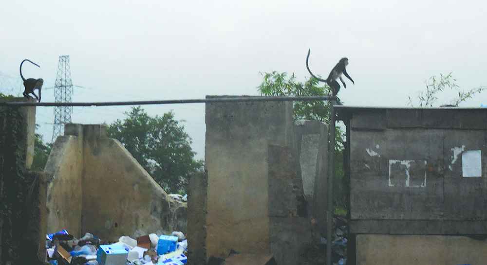
Mona monkeys scavenging around a refuse dump near the Lagos Lagoon.
The ELISA Kit used for the LASV NP antigen assay has been shown to have 95% sensitivity and 97% specificity when compared with standard quantitative polymerase chain reaction [16]. However, performance characteristics of the LASV NP IgG and IgM assays have not been published. Crossreactivity with closely related viruses is possible but, because recombinant proteins were used, it was likely minimal. There is, therefore, a critical need to broaden Lassa fever surveillance to include monkeys and other NHPs for their role in the epidemiology of Lassa fever.
FINANCIAL SUPPORT
This research received no specific grant from any funding agency.
CONFLICTS OF INTEREST
The authors have no conflicts of interest to declare.
ACKNOWLEDGMENTS
The author is grateful to the members of staff in the different wildlife facilities used in this study for their kind assistance in the animal restraint procedures.
REFERENCES
Cite this article
TY  - JOUR
AU  - Bamidele Nyemike Ogunro
AU  - Babasola Oluseyi Olugasa
AU  - Adeyemi Kayode
AU  - Olayinka Olabisi Ishola
AU  - Oluseyi Noah Kolawole
AU  - Eugene Amiewanlen Odigie
AU  - Christian Happi
PY  - 2019
DA  - 2019/05/01
TI  - Detection of Antibody and Antigen for Lassa Virus Nucleoprotein in Monkeys from Southern Nigeria
JO  - Journal of Epidemiology and Global Health
SP  - 125
EP  - 127
VL  - 9
IS  - 2
SN  - 2210-6014
UR  - https://doi.org/10.2991/jegh.k.190421.001
DO  - 10.2991/jegh.k.190421.001
ID  - Ogunro2019
ER  -