
P.07 The Progression of Left Ventricular Ejection Time in Simulated Microgravity
- DOI
- 10.2991/artres.k.201209.021How to use a DOI?
- Keywords
- Microgravity; pulse wave; LVET
- Abstract
Introduction: Microgravity in space is known to cause major alterations in the cardiovascular system. Left ventricular ejection time (LVET) can be measured by the time from the onset point of the pressure wave to the incisura of the dicrotic notch. The aim of this study was to simulate microgravity by head-down tilt bedrest (HDT) to examine changes in LVET in female and male subjects.
Methods: 24 healthy subjects (16 males and 8 females, height 176 ± 7 cm, weight 77 ± 6 kg, age 37 ± 10 years) were enrolled in a HDT study. The bed rest study applied strict –6° HDT for 60 days. Pulse wave measurements were taken using an oscillometric pressure cuff on the brachial artery. LVET index (LVETi) was calculated according to Weissler et al [1]. LVETis of different measurement times were compared using repeated measures ANOVA with post-hoc analysis using paired t-tests and Bonferroni correction.
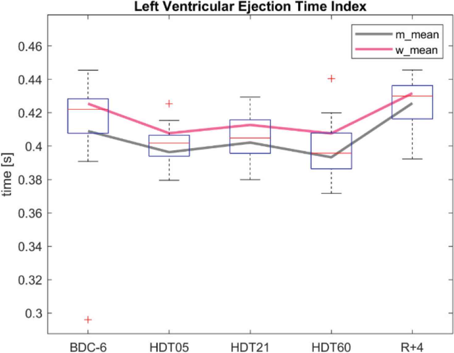
Results: Figure shows a decrease of LVETi during bed rest, followed by a sharp rise of LVETi after bed rest. Repeated measures ANOVA confirmed significant differences between measurement times. The increase of LVETi from each HDT measurement day to 4 days after HDT (R + 4) was significant (p < 0.001). There were no significant differences comparing male and female subjects.
 Figure
LVETi measured 6 days before (BDC-6), on the 5th day (HDT05), on the 21st day (HDT21), on the 60th day (HDT60), and 4 days after (R + 4) bedrest. Bold red and gray lines indicate female and male mean values.
Discussion/Conclusion: Overall, we conclude that LVETi decreased during HDT and reached four days after bed rest a similar level as before for both female and male subjects. As LVETi removes heart rate induced effects on LVET, the change in LVETi might be a result of change in ventricular ejection and afterload.
- Copyright
- © 2020 Association for Research into Arterial Structure and Physiology. Publishing services by Atlantis Press International B.V.
- Open Access
- This is an open access article distributed under the CC BY-NC 4.0 license (http://creativecommons.org/licenses/by-nc/4.0/).
Cite this article
TY  - JOUR
AU  - Stefan Orter
AU  - Stefan Möstl
AU  - Martin Bachler
AU  - Fabian Hoffmann
AU  - Christopher C. Mayer
AU  - Eugenijus Kaniusas
AU  - Michaela Reisinger
AU  - Siegfried Wassertheurer
AU  - Jens Tank
AU  - Bernhard Hametner
PY  - 2020
DA  - 2020/12/31
TI  - P.07 The Progression of Left Ventricular Ejection Time in Simulated Microgravity
JO  - Artery Research
SP  - S27
EP  - S27
VL  - 26
IS  - Supplement 1
SN  - 1876-4401
UR  - https://doi.org/10.2991/artres.k.201209.021
DO  - 10.2991/artres.k.201209.021
ID  - Orter2020
ER  -