
Multiple splenic artery aneurysms in a patient with idiopathic thrombocytopenic purpura: A case report, brief literature review and discussion
- DOI
- 10.1016/j.artres.2009.12.001How to use a DOI?
- Keywords
- Splenic; Abdominal; Multyple; Aneurysms; Thrombocytopenia; MDCT
- Abstract
Multiple splenic artery aneurysms in a patient with idiopathic thrombocytopenic purpura: A case report, brief literature review and discussion.
- Copyright
- © 2009 Association for Research into Arterial Structure and Physiology. Published by Elsevier B.V. All rights reserved.
- Open Access
- This is an open access article distributed under the CC BY-NC license.
Clinical case
A 69 year old female with a 10 year history of Idiopathic Thrombocytopenic Purpura (ITP) being treated with 60 mg of prednisone per day was admitted for worsening of her clinical condition and abdominal left upper quadrant pain. Contrast-enhanced computed axial tomography exam of her abdomen and pelvis revealed six calcified distal splenic aneurysms with partial luminal thrombosis (Fig. 1–4).
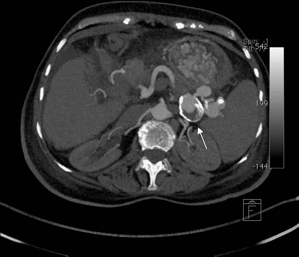
Contrast enhanced axial MIP image, in arterial phase, demonstrates several hilar splenic aneurysms. The largest and pointed one in this figure has an egg-shaped mural calcification and a partial luminal thrombosis.
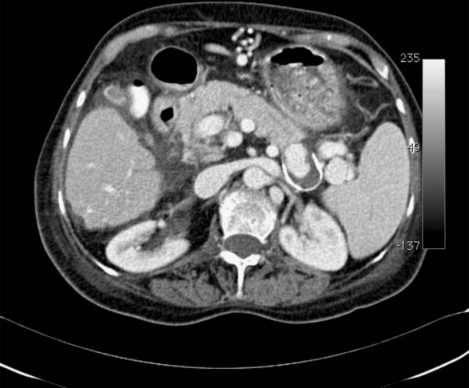
This contrast enhanced axial MIP image, in portal phase, also shows the hilar splenic aneurysms, still well opacified. This may mean a slow arterial flow.
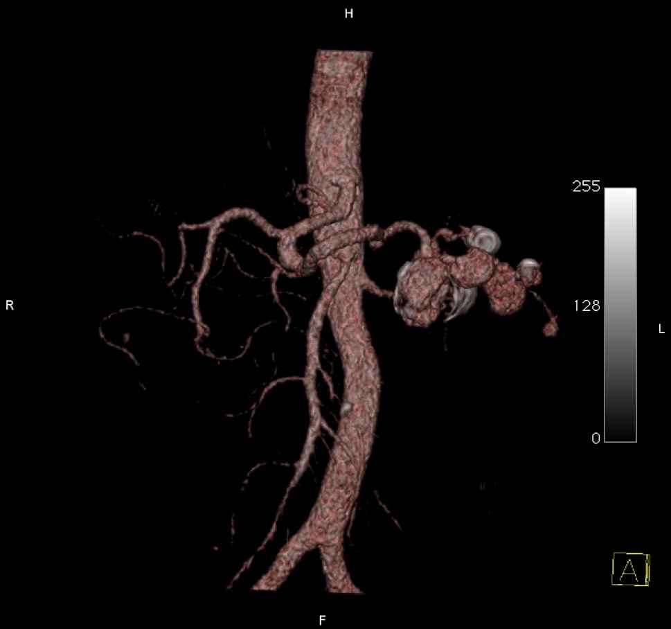
Coronal volumetric rendering (VRT) demonstrates multiple splenic aneurysms and their relation to the abdominal vasculature.
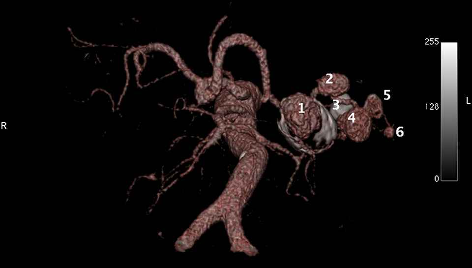
Inferior volumetric rendering (VRT) showing the splenic artery aneurysms’ location. Each one is numbered according to its position, from medial to lateral. Mural thrombi are seen in aneurysms marked as 1, 3 and 5.
Literature review and discussion
Although splenic artery aneurysms represents only 0.77% of all aneurysms3 it is a well known entity due to it’s risk of rupture, most commonly seen in young or pregnant females.3
The splenic artery is the third most common location of intra-abdominal aneurysms after abdominal aortic aneurysm and iliac artery aneurysm.4 It is four times more common in females but the risk of rupture is three times more common in males.8 Although its etiology is unknown, it is commonly associated with cirrhosis, hypertension, liver transplantation, and pregnancy. Less common associations exist with fibromuscular dysplasia, collagen vascular diseases, arteritis and alpha-1-antitrypsin deficiency.
Atheromatous disease is not considered a causative factor in splenic artery aneurysms.4,8
Splenic artery aneurysms have an average diameter of less than 3 cm, can be single or multiple, and are generally located in the distal portion of the artery although they can develop in any portion of the artery. They may display atherosclerotic changes, calcify, or thrombose. The majority of patients are asymptomatic until rupture, when they present with pain, hemodynamic instability, and internal hemorrhage.4,9
Several strategies exist to manage splenic artery aneurysms. Treatment should be considered, especially in patients with an increased risk of rupture such as those with liver transplants, cirrhosis, portal hypertension, pregnancy or young age. Nevertheless, there is currently no consensus on the treatment of asymptomatic patients. Factors to consider when planning management include the location of the lesion, age of the patient, associated surgical risks and the patient’s general health status.1,2,5–7
References
Cite this article
TY  - JOUR
AU  - David Busel
AU  - Alonso Yanez
AU  - Edgard Jimenez
AU  - Christopher K. Johansen
PY  - 2010
DA  - 2010/01/06
TI  - Multiple splenic artery aneurysms in a patient with idiopathic thrombocytopenic purpura: A case report, brief literature review and discussion
JO  - Artery Research
SP  - 24
EP  - 26
VL  - 4
IS  - 1
SN  - 1876-4401
UR  - https://doi.org/10.1016/j.artres.2009.12.001
DO  - 10.1016/j.artres.2009.12.001
ID  - Busel2010
ER  -