
Vasoreactivity of thoracic aorta in Nigella Sativa supplemented and/or exercise trained rats
- DOI
- 10.1016/j.artres.2017.12.006How to use a DOI?
- Keywords
- Nigella sativa; Exercise; Vascular sensitivity; Thoracic aorta
- Abstract
Background: Nigella Sativa (NS) induced vasorelaxation of aortic rings upon direct exposure has been documented in literature. This study was conducted to determine whether 8 weeks Nigella Sativa supplementation and/or exercise training may alter vascular sensitivity of rat aorta in normal, healthy rats.
Methods: In this Quasi Experimental Study, forty adult male Wistar albino rats were divided equally into 4 groups. One group served as the control (C), whereas the other three groups were Nigella-treated (N; 800 mg/kg Nigella daily orally via a feeding needle), Exercise-trained (Ex: 18 m/min speed, 2 h duration/day at 32 inclination) and Nigella-treated-exercise-trained (N-Ex: received both Nigella and exercise training). After 8 weeks, rats in all four groups were sacrificed. Their aortic rings were2 mounted in the organ bath. Tension in the aortic rings was measured with an isometric force transducer and recorded with a PowerLab data-acquisition system. Norepinephrine-induced aortic contractions in all four groups were compared by one-way analysis of variance (ANOVA).
Results: There were statistically insignificant differences in Norepinephrine-induced aortic contractions among all four groups (P value 0.26, 0.18, 0.07, 0.12, 0.58, 0.24, 0.06 with Norepinephrine 0, 10−7, 10−6, 10−5, 10−4, 10−3, 10−2 respectively). Also, no statistical difference was observed in term of body weight, heart weight, left ventricular weight and their indices in all four groups.
Conclusion: NS supplementation and exercise training alone or in combination; for 8 weeks’ duration, may not alter vascular reactivity in normal healthy rats.
- Copyright
- © 2018 Association for Research into Arterial Structure and Physiology. Published by Elsevier B.V. All rights reserved.
- Open Access
- This is an open access article distributed under the CC BY-NC license.
Introduction
Nigella Sativa (NS) or Black seed is a traditional miraculous medicinal plant. Many researches have revealed its extensive pharmacological effects including analgesic, antidiabetic, antitumor, immunomodulator, antimicrobial, anti-inflammatory, spasmolytic, bronchodilator, hepato-protective, renal protective, gastro-protective and antioxidant properties.1–4 Owing to its broad-range medicinal value, NS has been ranked among the top evidence based herbal medicines. Prophet Mohammad (PBUH) stated, “There is healing in Black Seed for all diseases except death”.5
NS extract has been shown to reduce blood pressure in experimentally hypertensive rats6,7 and hypertensive patients.8 Even in healthy volunteers, oral daily administration of 5 ml NS oil for 8 weeks lowered systolic and diastolic blood pressures (BP).9 Mechanism behind this NS-induced BP lowering effects is not well established. One possible justification might be the reduced systemic vascular resistance owing to decreased vasoconstrictor response to agents like Norepinephrine (NE). An in vitro study revealed vasorelaxant effects of NS seed extract on the aortic rings pre-contracted with Phenylephrine hydrochloride and Potassium Chloride.10 Niazmand et al. inferred that relaxation was produced by inhibition of Ca2+ channels and intracellular calcium release. Another in vitro study conducted by Cherkaoui-Tangi et al. reported inhibition of Ca2+-induced contractions in aortic rings pre-incubated with Nigella oil11 They suggested that this NS-induced vasodilation was endothelium-independent and from the blockade of both voltage-sensitive and receptor-operated calcium channels.
Exercise training improves cardiovascular health. With regular physical training, BP (systolic and diastolic both) has been significantly reduced in hypertensive subjects.12 Mechanism underpinning this post-exercise hypotension involves a drop in systemic vascular resistance brought about by both; neural and local factors.13–15 Recent studies conducted in obese rats to see the effects of acute and intermittent exercises on aortic vasoreactivity have shown exercise-induced attenuation of aortic contractions induced by Noradrenaline.16,17 The authors have attributed this to enhanced antioxidant enzyme activity and reduced free radical generating capability of exercise.
The documented vasorelaxatory effects of NS on aortic rings in in vitro studies, blood pressure lowering effects of NS and exercise training in in vivo, evidence of exercise-induced improved aortic vasoreactivity in obese rats led us to hypothesize that NS supplementation and/or exercise training may alter vascular sensitivity in normal, healthy rats.
The primary aim of the study was to find out the effects of 8 weeks’ period of exercise training, Nigella Sativa supplementation, or a combination of both on aortic vascular sensitivity to vasoconstrictor agents. As a secondary measure, we recorded indices of cardiac hypertrophy (heart weight, left ventricular weight, tibial length) as well in all groups.
Materials and methods
This research was carried at department of Physiology, College of Medicine, Imam Abdulrahman Bin Faisal University, Dammam after approval by the Institutional Review Board. The Animal welfare guidelines and research protocols were strictly adhered to.
Forty normal adult Wistar albino male rats (weighing 150–250 g), obtained from university animal house, were housed individually in labeled cages with adequate ventilation, illumination, laboratory chow feed and water ad libitum.
Rats were randomly divided into four equal groups (n = 10): Nigella-treated (N) group, exercise-trained (Ex) group, Nigella-treated–exercise-trained (N-Ex) group, and a control (C) group. Rats of the N group were given 800 mg/kg Nigella (10 g freshly ground Nigella/100 ml distilled water) daily at around 11 am for 8 weeks, via oral gavage. Rats of the C group received vehicle (distilled water). Rats of Ex group were trained on a treadmill (IITC life science, 5 lane rat treadmill, Victory Blvd Woodland Hills, CA) daily for 8 weeks. Speed, grade, and duration were gradually increased during the first week till final protocol of 18 m/min speed, 32 inclination, for a 2 h/session was attained.18,19 An electric grid (0.2 mA) at the back of the belt discouraged rats from discontinuing. Rats of the N-Ex group received both experimental interventions, i.e. Nigella supplementation and exercise training.
At the end of the 8-week protocol, rats were anesthetized with a ketamine cocktail (60% ketamine, 40% xylazine; 0.8 ml/kg body weight intra-peritoneally). Hearts were extracted through longitudinal abdominal incisions and soaked in cold Ringer’s solution. After removing excess of connective tissue, lumens were rinsed with Ringer’s solution, the hearts were blotted dry and weighed. Left ventricles were weighed after removing right ventricles and both atria.
One leg was removed above the knee joint, and the length of the tibia from the condyles to the tip of the medial malleolus was recorded.
The thoracic aorta was immersed in ice-cold Krebs-Henseleit (KH) solution (pH 7.4) containing (mMol/L): NaCl 118.4, KCl 4.7, CaCl2 2.52, MgSO4 1.18, KH2PO4 1.18, NaHCO3 25.0, and
The dissected aorta was cut into 1.8 mm long ring segments and mounted onto two stainless-steel stirrups. The lower stirrup was anchored to the tissue mounting hook, and the upper stirrup related to a thread to the isometric force transducer for tension measurement and recording with a PowerLab data-acquisition system. The aortic rings were then immersed in a 20 ml organ bath containing KH buffer continuously bubbled with 95% O2 and 5% CO2 and maintained at 36.5 °C.
The organ bath used was ML0146 (AD Instruments) equipped with four tissue chambers, preheating reservoir coils, gas diffusers, tissue holders, micro positioners, a water pump, and thermostat controller. Tissue responses were recorded using force transducer MLT0201, Quad bridge amplifier FE224, and PowerLab® PL3508 data-acquisition system with LabChart Pro®.
The rings were stretched to a resting tension of 1 g and equilibrated for at least 1 h, with bath fluid (KH) being changed every 15 min.20 Baseline tension was recorded after calibration. Aortic rings were then exposed to varying strengths of norepinephrine (NE) starting from low concentration to high concentration, i.e., 10−7–10−2. The contraction response was defined as an increase in tension from the resting value after equilibration.
All chemicals were purchased from the Sigma pharmaceutical company. Strengths prepared were 10−3, 10−4, 10−5, 10−6, and 10−7. A stock solution of norepinephrine (10−2) was made with deionized water (90%), HCl (10% to ensure solubility), and ethylenediaminetetraacetic acid (ethylenediaminetetraacetic acid; 100 μg/1 ml to prevent oxidation of NE). All the drugs were serially diluted in distilled water before each experiment. The concentration of a drug is expressed as the final concentration in the bath solution.
Maximum response was calculated for all doses through LabChart®. Statistical analysis was carried out by IBM SPSS statistics version 20. A one-way analysis of variance (ANOVA) was used to determine the significance of differences among NE-induced contractions among all four groups. All values were presented as Means ± Standard Error of Mean. P < 0.05 was required for significance.
Results
Mean values for different doses of NE (10−7–10−2) on intact aortic rings from all four groups were compared. Being a vasoconstrictor agonist, exposure of aortic rings to varying concentrations of NE caused them to contract in a dose dependent manner. NS-fed or exercise-trained rats, exhibited statistically insignificant differences among NE-induced aortic constrictions compared to controls. Even the combination of both procedures (i.e. NS supplementation and exercise training) failed to produce any significant synergistic/potentiating effects in diminishing contractile responsiveness (Table 1 and Fig. 1). Also, no statistical difference was observed in term of body weight, heart weight, left ventricular weight and their indices in all four groups (Table 2).
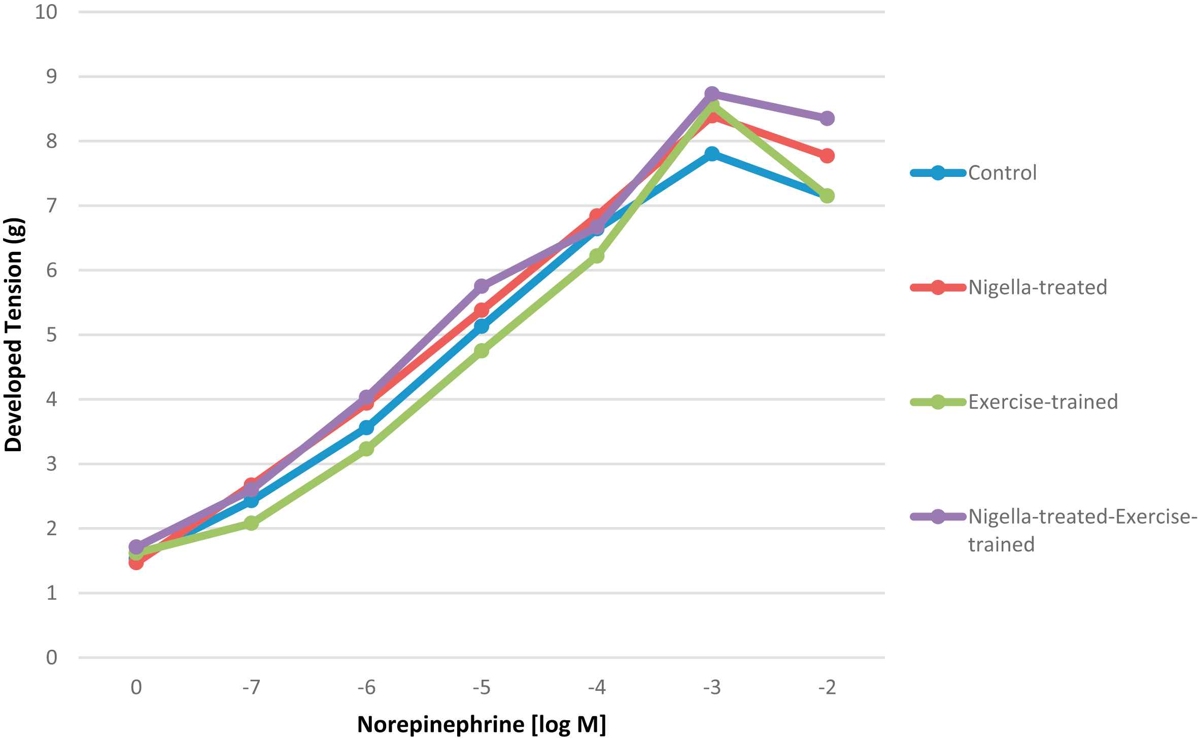
Tension (g) developed in all four groups after exposure to varying concentration of Norepinephrine.
| Group | Developed Tension (g) (Mean ± SEM) | ||||||
|---|---|---|---|---|---|---|---|
| Baseline | NE 10−7 | NE 10−6 | NE 10−5 | NE 10−4 | NE 10−3 | NE 10−2 | |
| Control | 1.53 ± 0.07 | 2.43 ± 0.27 | 3.56 ± 0.28 | 5.13 ± 0.38 | 6.64 ± 0.37 | 7.80 ± 0.47 | 7.15 ± 0.37 |
| Nigella treated | 1.47 ± 0.11 | 2.67 ± 0.20 | 3.94 ± 0.23 | 5.38 ± 0.32 | 6.84 ± 0.27 | 8.39 ± 0.33 | 7.77 ± 0.39 |
| Exercise | 1.62 ± 0.10 | 2.08 ± 0.20 | 3.23 ± 0.24 | 4.75 ± 0.20 | 6.22 ± 0.38 | 8.56 ± 0.32 | 7.15 ± 0.37 |
| N-Ex | 1.71 ± 0.07 | 2.60 ± 0.09 | 4.03 ± 0.15 | 5.75 ± 0.22 | 6.67 ± 0.25 | 8.73 ± 0.18 | 8.35 ± 0.29 |
| P value (ANOVA) | 0.26 | 0.18 | 0.07 | 0.12 | 0.58 | 0.24 | 0.06 |
NE = Norepinephrine: N-Ex = Nigella-treated-exercise-trained.
Comparison (ANOVA) of Tension (g) developed in all four groups after exposure to varying concentration of Norepinephrine.
| Control | N | Ex | N-Ex | P value | |
|---|---|---|---|---|---|
| Initial body weight | 249.6 ± 21.7 | 207.5 ± 62.0 | 237.4 ± 14.7 | 234.3 ± 35.9 | 0.40 |
| Final body weight | 272.4 ± 30.0 | 248.8 ± 63.0 | 269.4 ± 22.0 | 218.3 ± 47.8 | 0.24 |
| HW (g) | 0.921 ± 0.151 | 0.750 ± 0.109 | 0.946 ± 0.057 | 0.831 ± 0.198 | 0.17 |
| LVW (g) | 0.686 ± 0.638 | 0.622 ± 0.157 | 0.703 ± 0.072 | 0.616 ± 0.121 | 0.55 |
| HW/BW (×103) | 3.4 ± 0.4 | 3.2 ± 0.9 | 3.5 ± 0.2 | 3.8 ± 0.7 | 0.41 |
| LVW/BW (×103) | 2.0 ± 1.1 | 2.5 ± 0.2 | 2.6 ± 0.2 | 2.9 ± 0.5 | 0.22 |
| LVW/HW | 0.566 ± 0.317 | 0.850 ± 0.296 | 0.742 ± 0.039 | 0.746 ± 0.033 | 0.31 |
| HW/TL | 0.022 ± 0.004 | 0.018 ± 0.003 | 0.021 ± 0.002 | 0.020 ± 0.004 | 0.36 |
| LVW/TL | 0.013 ± 0.007 | 0.015 ± 0.003 | 0.016 ± 0.002 | 0.015 ± 0.002 | 0.77 |
HW: Heart weight, LVW: Left ventricular weight, BW: Body weight, TL: Tibial length.
N: Nigella-treated group.
Ex: Exercise-trained group.
N-Ex: Nigella-treated–exercise-trained group.
Comparison of rat body weights, heart weight, left ventricular weight, and their indices among all four groups.
Discussion
Vasoreactivity refers to acute changes in vascular function. It reflects arterial vascular functional state and is considered as a prognostic indicator of arterial health. Enhanced vasoreactivity may be an early indication for the development of cardiovascular diseases.21
The main findings of the present study lie in the fact that 8 weeks NS supplementation and exercise training alone or in combination failed to produce any significant alteration in vascular reactivity of normal rats.
Our study results are in contrast to Abbasnezhad et al.,22 who showed that 8 week supplementation of NS seed extract significantly improved aortic reactivity to vasoconstrictor and vasodilator agents in Streptozotocin-induced diabetic rats. The reason for results discrepancy might be the use of pure NS extract by Abbasnezhad et al., compared to mere suspension in our study; as pure extract is more concentrated compared to suspension. Another possibility could be the subject variation; i.e. diabetic rats versus normal rats. It has been shown that diabetes leads to endothelial dysfunctions by several metabolic derangements, including the hyperglycemia, hyperlipidemia and oxidative stress.23 NS treatment can produce significant hypoglycemic, hypolipidemic and antioxidant effects in diabetes.24,25 Therefore, improved aortic reactivity to vasoactive agents in diabetes with concurrent administration of NS might be, in part or completely, due to NS’s hypoglycemic, hypolipidemic and antioxidant effects; rather than its direct effects. Normal rats in whom metabolic derangements (hyperglycemia, hyperlipidemia, oxidative stress) and/or endothelial dysfunctions are absent, 8 weeks’ supplementation with NS might not cause an additive advantage on vasoactivity.
Our study results are contrary to Cherkaoui-Tangi et al.11 as well. In that study, pre-incubation of the rings with Nigella oil effectively antagonized Ca2+ -induced contractions, leading authors to believe that Nigella oil may interfere with both voltage and receptor-operated Ca2+ channels. In another study,10 researchers demonstrated that direct exposure of rat aortic rings to NS reduced the contraction induced by Potassium chloride. The disparity could be due to the use of NS pure extract vs suspension; secondly in vitro vs in vivo. Researchers all over the globe agree that in vitro responses may not be identical to in situ or in vivo preparations and results from an in vitro/ex vivo study cannot be extrapolated to in vivo because in vitro tissues are studied under controlled conditions without extrinsic neural factors, circulating hormones, blood flow and shear stress etc. Hence, in vitro measurements only help to anticipate what can happen, not what does happen in integrative and intricate in vivo conditions.
A secondary purpose of this study was to determine that whether 8 weeks’ exercise training induces a change in responsiveness of rat aortas to adrenergic stimulation. In the current study, exercise training had no effect on aortic vasoreactivity, verifying prior researches which documented that unless the total volume of exercise training is very high, significant changes in aortic vasoreactivity are unlikely to occur. Our findings support the studies26,27 in which no significant difference was found in aortic vasoreactivity between rats of the control group and rats subjected to 4 weeks swimming exercise OR 1-, 2-, 4- and 10- weeks treadmill exercise training respectively.
However, our study results differ from those reported by Howard et al.,28 The discrepancy between our results and Howard can be justified based on different experimental design, the intensity of the exercise, or the species of animal used in the study. For example, Howard protocol was a single bout of exercise consisting of 24 m/min treadmill running until exhaustion (an average exercise duration of 16 min) in normotensive rabbits whereas the protocol in the present study was 8 weeks training of treadmill running at 18 m/min for 2 h/session in rats. These variations in relative exercise intensity and duration might be the reason for disparity in results. Our study results differ from Xu T et al.,16 as well who reported a significant decrease in Noradrenaline induced aortic contractions in rats subjected to swimming exercise (30 min/time, 3 times/day, 5 days/week for 8 weeks, moderate intensity) with loads of 5% of their body weight attached to their tails. Xu T rats received intraperitoneal anaesthesia with 20% urethane (0.5 ml/kg), whereas rats in our study were anesthetized with ketamine cocktail. Urethane is believed to influence multiple neurotransmitter systems at an aesthetic concentration.29 This may account for results disparity.
In the end, even the group exposed to exercise training and NS supplementation both, failed to produce any significant effect on aortic vasoreactivity compared to rest of the groups.
Briefly, we conclude that NS supplementation and exercise training alone or in combination for 8 weeks may not produce any significant effect on vascular reactivity when endothelial functions are normal.
Limitations
Use of mere suspension of Nigella Sativa instead of the pure extract, limited duration and/or amount of Nigella Sativa supplementation/exercise training are some of the limitations in this study. Measurement of Tail pressures could not be performed due to generation of unnecessary oscillations/signals from the device.
Conflict of interest
None.
References
Cite this article
TY  - JOUR
AU  - Rabia Latif
AU  - Al-Asoom LI
PY  - 2018
DA  - 2018/01/11
TI  - Vasoreactivity of thoracic aorta in Nigella Sativa supplemented and/or exercise trained rats
JO  - Artery Research
SP  - 38
EP  - 42
VL  - 21
IS  - C
SN  - 1876-4401
UR  - https://doi.org/10.1016/j.artres.2017.12.006
DO  - 10.1016/j.artres.2017.12.006
ID  - Latif2018
ER  -