
P2.20 ACOUSTIC LOCALISATION OF CORONARY ARTERY STENOSIS: WAVE PROPAGATION IN SOFT TISSUE MIMICKING GELS
- DOI
- 10.1016/j.artres.2013.10.081How to use a DOI?
- Abstract
Background: Turbulent flow downstream of atherosclerotic plaques produces low amplitude shear waves which travel through the chest and can be measured by skin sensors. This acoustic signature may provide a cheap non-invasive way to diagnose arterial disease. We report measurements of shearing oscillations and flow-induced turbulence in soft tissue-mimicking gels which provide input to a numerical model of soft tissue behaviour described in a companion presentation.
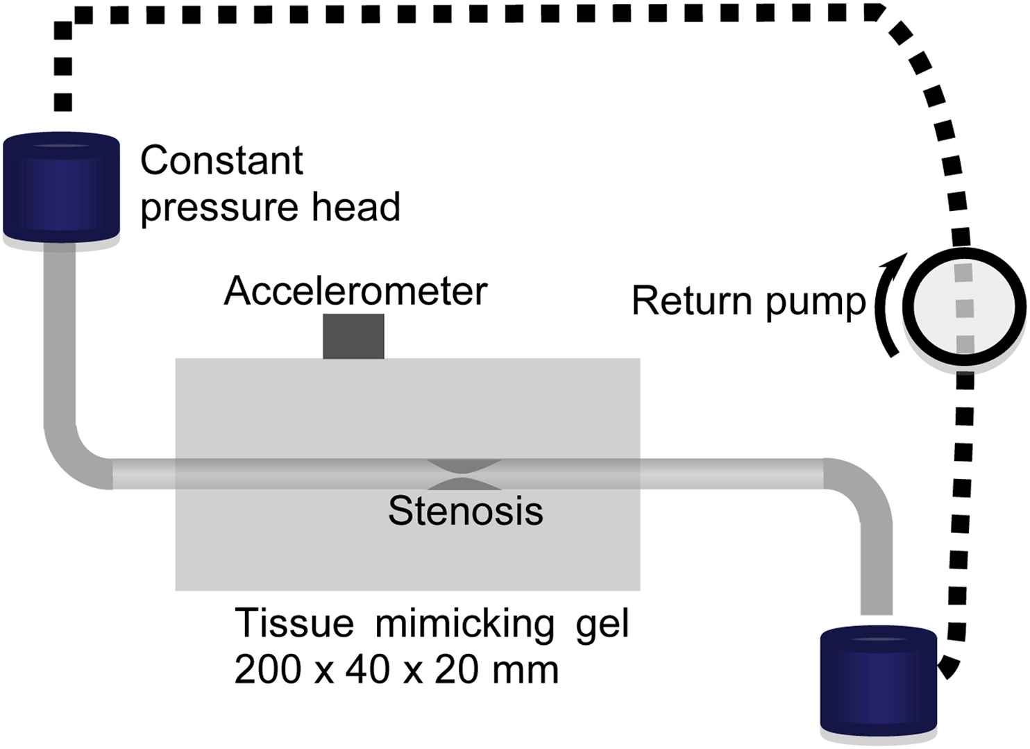
Methods: Cylindrical specimens of 3% agarose gel were cast around an axial rod and bead connected to an electromechanical vibrator (figure 1), to generate shear-waves of known characteristics and location (frequency 250–750 Hz, amplitude 10–50 μm). Displacement was mapped optically by tracking the movement of carborundum particles on the surface. In the flow study (figure 2) a silicone rubber tube (i.d. 4.5mm) containing a stenosis was embedded in a cuboidal gel phantom and lateral displacement of the gel surface was mapped by a piezo-electric accelerometer.
Results: Forced oscillations produced movement in the same direction at the gel surface, amplitude 10–50% of the bead’s movement. Amplitude modulation (≈5%) at around 40Hz, probably due to resonance in the gel, was also seen. Lateral movement (200–800Hz) of the gel surface caused by flow-induced turbulence increased monotonically with turbulence magnitude.
Conclusions: The methods described above provide internally consistent and repeatable data, validating the numerical models. The next steps will compare computational results with measurements in progressively more realistic representations of the chest aiming ultimately to produce a device suitable for screening/diagnosis of coronary artery disease.
Figure. 1. Forced vibration rig. Gels cast with bead in various positions. Laser measures bead movement; camera measures surface movement
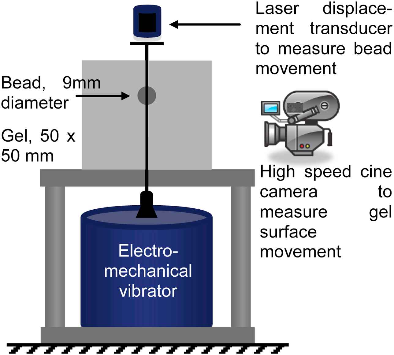
Figure 2. Steady flow rig. Measurements made at various flow rates, tube depths and stenosis severity. Accelerometer position varied.
- Open Access
- This is an open access article distributed under the CC BY-NC license.
Cite this article
TY  - JOUR
AU  - S.E. Greenwald
AU  - H.T. Banks
AU  - M.J. Birch
AU  - M.P. Brewin
AU  - S. Hu
AU  - Z.R. Kenz
AU  - C. Kruse
AU  - D. Mehta
AU  - J. Reeves
AU  - S. Shaw
AU  - J.R. Whiteman
PY  - 2013
DA  - 2013/11/11
TI  - P2.20 ACOUSTIC LOCALISATION OF CORONARY ARTERY STENOSIS: WAVE PROPAGATION IN SOFT TISSUE MIMICKING GELS
JO  - Artery Research
SP  - 124
EP  - 124
VL  - 7
IS  - 3-4
SN  - 1876-4401
UR  - https://doi.org/10.1016/j.artres.2013.10.081
DO  - 10.1016/j.artres.2013.10.081
ID  - Greenwald2013
ER  -