
P85 HIGH FRAME RATE DYNAMIC DISPLAY ULTRASOUND VECTOR FLOW IMAGING FOR QUANTITATIVE STUDIES OF HEMODYNAMICS OF CAROTID ARTERIES
- DOI
- 10.1016/j.artres.2017.10.101How to use a DOI?
- Abstract
Advanced atherosclerotic patients are faced with significant risks of stroke, which are very likely to cause death or irreversible physical disability. However, the growth of artery stenosis usually needs a very long development. Early diagnosis is necessary and requires detailed and accurate quantitative hemodynamics to be supported. The paper proposes an angle-independent ultrasound flow imaging technique for carotid arteries, which allows true velocity vectors measurement, obtaining both value and direction of blood flow.
The proposed vector flow imaging is implemented based on multi-directional Doppler interleaved transmission [1,2], with high frame rate dynamic display [1] and zone sonography technology [3].
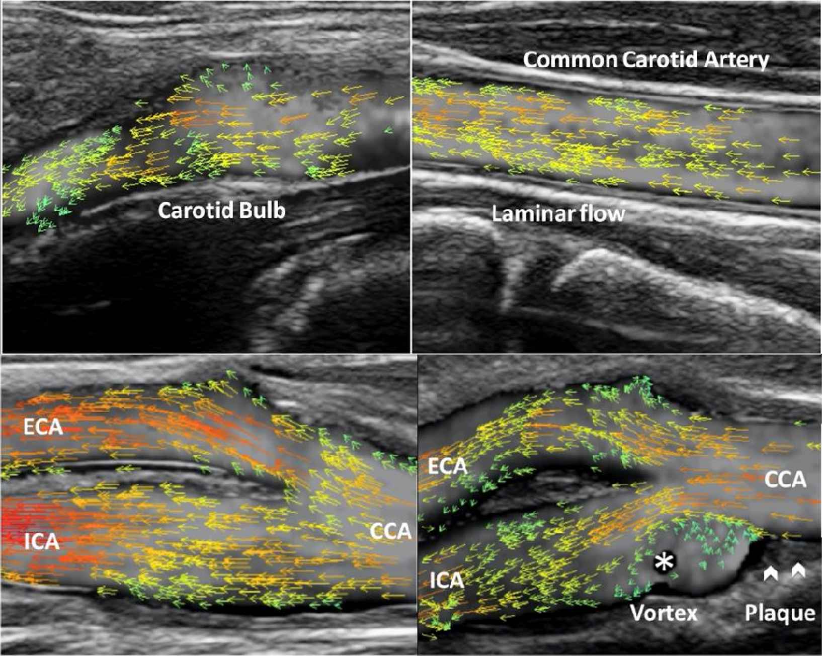
Hemodynamics becomes extremely complicated when plaques develop in the carotid bulb. The dynamic display with velocity vectors assesses flow patterns, e.g. laminar flow, vortex and turbulence (Examples are shown in the figure). The circular variance for the angles of vectors in a desired region of interest can be calculated, allowing disturbance quantification for the non-laminar flow. The method is capable of measuring volume flow (VF) and wall shear stress (WSS) at different locations. To ensure the accuracy both VF and WSS are calculated based on a frame rate of 400–600 Hz and vector velocities.
The high frame rate vector flow imaging has been implemented in a commercial ultrasound system. It provides various quantitative results such as circular variance, VF and WSS, which are useful for hemodynamics studies of complex flow. This could make the early prevention and diagnosis of carotid disease possible.
- Open Access
- This is an open access article distributed under the CC BY-NC license.
References
Cite this article
TY  - JOUR
AU  - Yigang Du
AU  - Xujin He
AU  - Yingying Shen
AU  - Lei Zhu
AU  - Alfredo Goddi
PY  - 2017
DA  - 2017/12/06
TI  - P85 HIGH FRAME RATE DYNAMIC DISPLAY ULTRASOUND VECTOR FLOW IMAGING FOR QUANTITATIVE STUDIES OF HEMODYNAMICS OF CAROTID ARTERIES
JO  - Artery Research
SP  - 77
EP  - 77
VL  - 20
IS  - C
SN  - 1876-4401
UR  - https://doi.org/10.1016/j.artres.2017.10.101
DO  - 10.1016/j.artres.2017.10.101
ID  - Du2017
ER  -