
A convenient three dimensional model to teach the arterial supply of the brainstem
- DOI
- 10.1016/j.artres.2009.03.001How to use a DOI?
- Keywords
- Artery; Brainstem; Anatomical education
- Abstract
An easier approach of teaching the three dimensional nature of arteries of the brainstem is being proposed and aids the understanding of clinical vascular conditions of the brain. Various regions of a standing student appear to represent different parts of the brainstem: head (midbrain), trunk (pons), thigh (medulla oblongata), leg (spinal cord), satchel (cerebellum) and plastic box (fourth ventricle). The vertebral arteries travel proximal to the spinal cord and medulla oblongata and unite at the position of the belt buckle to form the basilar artery. The basilar artery runs superiorly and finally bifurcates laterally at the neck of the student, to form the posterior cerebral artery. The teaching aid is simple, convenient and depicts 19 arteries of brainstem and circle of Willis.
- Copyright
- © 2009 Association for Research into Arterial Structure and Physiology. Published by Elsevier B.V. All rights reserved.
- Open Access
- This is an open access article distributed under the CC BY-NC license.
An easier approach of teaching the three dimensional nature of arteries of the brainstem is being proposed and aids the understanding of clinical vascular conditions of the brain Figs. 1 and 2. Various regions of a standing student appear to represent different parts of the brainstem: head (midbrain), trunk (pons), thigh (medulla oblongata), leg (spinal cord), satchel (cerebellum) and plastic box (fourth ventricle). The vertebral arteries travel proximal to the spinal cord and medulla oblongata and unite at the position of the belt buckle to form the basilar artery. The basilar artery runs superiorly and finally bifurcates laterally at the neck of the student, to form the posterior cerebral artery. The teaching aid is simple, convenient and depicts 19 arteries of brainstem and circle of Willis.
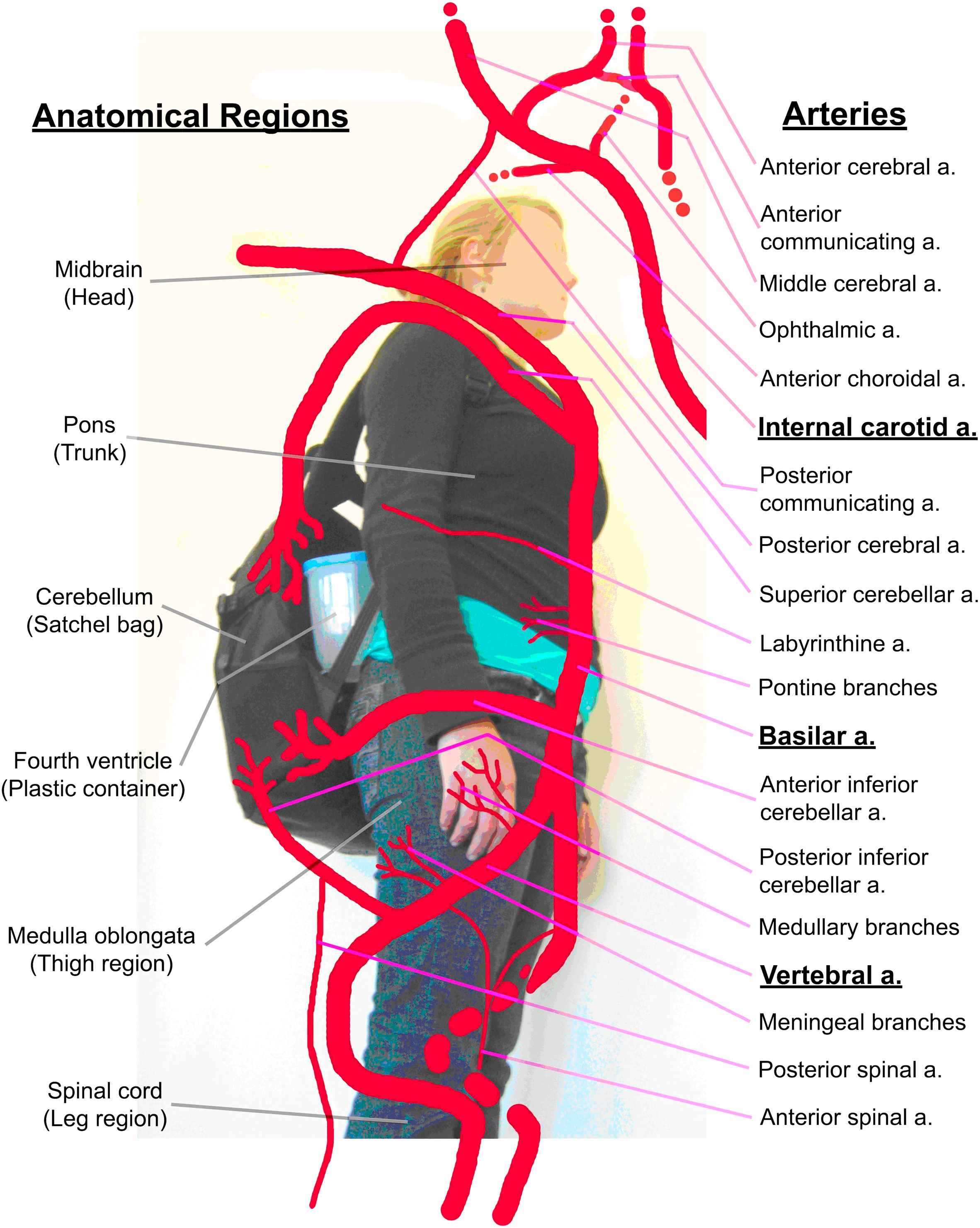
The proposed model for teaching the arterial supply of the brainstem.
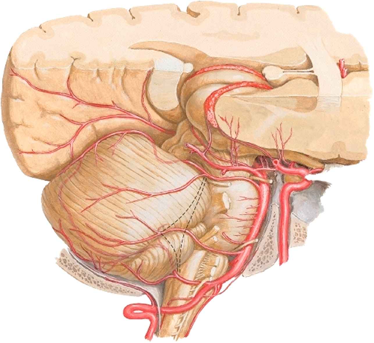
An anatomical illustration of the arterial supply of the brainstem from ‘An Atlas of Human Anatomy’ 3rd Edition by Frank Netter, Saunders–Elsevier (2003).
Cite this article
TY  - JOUR
AU  - H. Gangata
PY  - 2009
DA  - 2009/04/21
TI  - A convenient three dimensional model to teach the arterial supply of the brainstem
JO  - Artery Research
SP  - 89
EP  - 90
VL  - 3
IS  - 2
SN  - 1876-4401
UR  - https://doi.org/10.1016/j.artres.2009.03.001
DO  - 10.1016/j.artres.2009.03.001
ID  - Gangata2009
ER  -