
Relationship between the aortic valves and an anatomical landmark using chest CT scan
- DOI
- 10.1016/j.artres.2011.09.001How to use a DOI?
- Keywords
- Aortic arch; Aortic valves; Height; Hyoid bone; Morphometry
- Abstract
Purpose: In order to determine if the height of a subject could be a reliable surrogate variable to determine the pulse wave travelling distance within the aorta, we investigated the anatomical distance between the aortic valve nidus and the hyoid bone.
Methods: Using 28 patient’s chest CT-scans. From Multiplan reconstructed oblique cuts we measured 1) the length of the aortic arch from the aortic valve (AV) to the intercept of a horizontal line passing through the aortic valves and crossing the descending aorta at mark HD (see figure), and 2) the distance between the HD mark to the Hyoid Bone (HB).
Results: There was a correlation between the AV-HD distance and the HD-HB distance (non-parametric r = 0.66, p < 0.0001) and the AV-HD distance were positively correlated to the height of the subjects (r = 0.60, p < 0.002).
Conclusions: 1- The AV-HD distance projects to a constant anatomical landmark (i.e. the hyoid bone) 2- The size of this arterial segment is significantly correlated to the height of the subjects. These preliminary results could be useful for a more accurate determination of the pulse wave velocity.
- Copyright
- © 2011 Association for Research into Arterial Structure and Physiology. Published by Elsevier B.V. All rights reserved.
- Open Access
- This is an open access article distributed under the CC BY-NC license.
Introduction
Techniques to measure pulse wave velocity (PWV) are of increasing interest. It is the recognized indice of the arterial stiffness. “The gold standard” is represented by carotid-to-femoral pulse wave velocity. With this technique, to assess PWV, we divide the pulse travelled distance by its travel time. Therefore this path length is of crucial importance and has been largely debated.1
The aim of this work was to support the fact that the aortic valves (AV) can be related to a stable landmark. And second, we sought to determine whether a relationship exists between the length of the arterial system and the height of individuals.
Material and methods: We included 28 thoracic CT scans (General Electric, Bright speed, v4.3) of 19 men (range 41–90 years; median 66) and 9 women (50–98 years; 62 years). We measured their height and weight and all subjects gave their consent. The study was approved by the local ethic committee (CME) of Redon Hospital. We used the JMP 7.0 software (SAS Institute Inc. NC-USA) for statistical analysis, and non-parametric spearman’s R and the least squares regression were calculated.
Measurements on the CT scans were performed by a trained radiologist. The examinations were performed in patients referred to the radiology department of Redon hospital between March and April 2009 for a standard chest CT scan, regardless of their primary pathology. Patients were in supine resting position during the examination. MultiPlan Reconstruction oblique scan views were made after identification of the aortic valves (AV) in the horizontal section. The aortic arch was measured from the aortic valves (AV) to the intersection with the horizontal line passing through the aortic valves and the thoracic descending aorta (HD - Fig. 1). From this latest point (HD), we measured the distance to the hyoid bone (HD-HB).
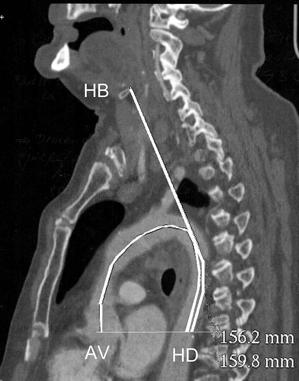
Measurement in semicircle of the aortic arch (AV-HD) = 156.2 mm, and 159.8 mm distance to the hyoid bone (HD-HB).
Results: The half-circular aortic segment measured between AV and HD was correlated to the distance to the hyoid bone (i.e. between HD and HB – see Fig. 1) : R = 0.66; p < 10 − 3, y = 0.95x + 10.56 (Fig. 2).
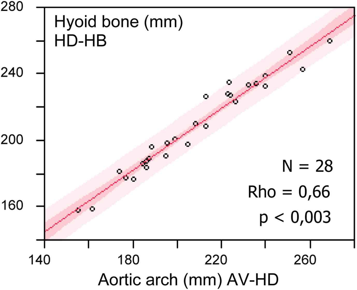
Correlation between the extent of the aortic arch and the distance to the hyoid bone. Y = 0.94X + 14.23.
The aortic segment AV-HD was also proportional to the height of individuals (R = 0.60; p < 0.002; y = 1.8x–83.12). There was no relationship with weight (R = 0.28; p = 0.24), nor with age and these distances (R = 0.32; p = 0.10), neither with sex.
Discussion: Although the relationship between the length of the arterial system and height is intuitive, we wanted to confirm it using CT scann. Vermeersh et coll have already reported that the travelled distance from carotid to femoral is also related to body height.2
Tsuguo Igari3 studied the length of the entire aorta from the subclavian artery to the renal artery based on computed tomography (CT images) and showed a positive correlation between height and the length of the entire aorta.
Although we did not found such relationship, Sugawara4 studied the length of the ascending aorta using MRI in 256 apparently healthy subjects and reported a strong relationship between age and the length of this segment, but not with the descending aorta.
Lantelme5 et al studied the relationship between the prognostic value of the PWV and the way to measure the travel distance from carotid to femoral measurement site. They concluded that PWV is more predictive of cardiovascular events when the distance D is estimated from the body height as Weber et coll have suggested this distance based on body height (D = height/4 + 7.28); as well as Filipovsky et al proposed to use algorithms based on body height to estimate the pulse travelled distance. According to Weber6 et coll, the best agreement with invasive measurements was found for the method of subtracting carotid-suprasternal notch distance from suprasternal notch-femoral distance.
Recently, Hybrechts7 demonstrated that the carotid to femoral artery distance (measured by tape) multiplied by 0.8, yielded the best agreement with the reference aortic path length measured with MRI in 98 healthy subjects.
We support the fact that the aortic valves represent the origin point for the pulse wave. In our work, valves project in front of the hyoid bone when the aorta is virtually unrolled. The aortic valves nidus is related to a stable anatomical landmark i.e. the Hyoid bone. This finding could be of interest to assess the pulse wave velocity, even of the travel path then for the travel time.
Conclusion: The unrolled aortic arch virtually project on the hyoid bone. Our data are of interest in the non-invasive determination of the length of the travelling carotid-femoral pulse wave distance and may be taken into account in the evaluation of the transit time. Several authors have deducted the aortic length from the height, or from a constant value of the direct measurement of the carotid to femoral distance over the body. If we admit that the aortic arch is proportional to height, then we may admit that the whole arterial system may be proportional to height even from the aortic valves to the extremities as finger or toe. Even if this hypothesis may be intuitive, it needs to be proven.
References
Cite this article
TY  - JOUR
AU  - Magid Hallab
AU  - Pascal Chevalet
AU  - Amine Dahou
AU  - Gilles Berrut
PY  - 2011
DA  - 2011/10/04
TI  - Relationship between the aortic valves and an anatomical landmark using chest CT scan
JO  - Artery Research
SP  - 55
EP  - 57
VL  - 6
IS  - 1
SN  - 1876-4401
UR  - https://doi.org/10.1016/j.artres.2011.09.001
DO  - 10.1016/j.artres.2011.09.001
ID  - Hallab2011
ER  -