
Carotid artery stiffness and cerebral pulsatility in children
- DOI
- 10.1016/j.artres.2018.05.002How to use a DOI?
- Keywords
- Vascular stiffness; Blood pressure; Hemodynamics; Carotid arteries; Blood vessels
- Copyright
- © 2018 Association for Research into Arterial Structure and Physiology. Published by Elsevier B.V. All rights reserved.
- Open Access
- This is an open access article distributed under the CC BY-NC license.
Compliance of the large arteries (e.g. the aorta and carotid artery) serve to dampen fluctuations in pressure and flow generated during a cardiac cycle, facilitating continuous target organ perfusion. Large artery stiffness increases transmission of hemodynamic pulsatility to the brain which causes cerebrovascular damage, cognitive decline and stroke later in life.1–3 To date, studies have primarily examined associations between large artery stiffness and cerebral pulsatility in adults.
Arterial stiffness increases from childhood to adolescence with some modification occurring through typical growth/development and some through subclinical atherosclerotic cardiovascular disease.4 As such, cerebral hemodynamic pulsatility may be influenced by the structural and/or functional status of large arteries at a much younger age than previously appreciated. The purpose of this study was to examine associations between large extracranial artery stiffness (aortic and carotid stiffness), extracranial hemodynamic pulsatility (carotid pulse pressure, flow pulsatility and pressure-flow wave intensity) and intracranial hemodynamic pulsatility (middle cerebral artery flow pulsatility) in children.
Methods
59 healthy children between 9 and 12 years of age (57% African American; 37% female) from the Syracuse City community participated in this study as part of the Environmental Exposures and Child Health Outcomes (EECHO) study. Detailed inclusion/exclusion criteria have been previously reported.5 This study was approved by the institutional review board of Syracuse University and SUNY Upstate University. All guardians and participants gave written consent and assent, respectively, prior to enrollment.
Applanation tonometry (Sphygmocor, AtCor Medical, Sydney, Australia) was used to acquire pressure waves at the common carotid and femoral pulse sites and used to calculate a pulse wave velocity (PWV).4 Brachial blood pressure was acquired using an oscillometric cuff (Mobil-o-graph, IEM, Germany). Carotid pressure waveforms were calibrated against brachial mean and diastolic blood pressure (BP) and used to estimate carotid systolic pressure, pulse pressure and augmentation index. The common carotid artery was imaged below the carotid bulb using Doppler-ultrasound (ProSound α7, Aloka, Tokyo, Japan) and a 7.5–10.0 MHz linear-array probe. Distension and flow velocity waveforms were used to estimate a single-point carotid PWV and carotid flow velocity pulsatility index (PI).6 Wave intensity analysis was used to derive a forward compression wave (W1) and backward travelling compression waves (negative area, NA).6 Middle cerebral artery (MCA) blood flow velocity was assessed using a 2-mHz transcranial Doppler (TCD) ultrasound probe (DWL Doppler Box-X, Compumedics, Germany) and used to derive a flow velocity PI.6
All data are reported as mean ± standard deviation. Associations of interest were explored using Pearson Correlation Coefficients. Path analysis was used to generate a model linking key vascular-hemodynamic constructs. Model fit was assessed using the normal fit index, the comparative fit index, the Chi-square minimum statistic, and the root mean square error of approximation. All analyses were performed using Statistical Package for the Social Sciences (SPSS, version 23, Chicago, IL) with significance set a priori as p < 0.05.
Results
Group characteristics are presented in Table 1. Systemic vascular-hemodynamic properties are shown in Table 2. Table 3 presents a correlation matrix of all vascular-hemodynamic properties examined in this study. There were significant inter-associations between carotid PWB-β, carotid PP, W1, NA, carotid PI, and MCA PI (Table 3, p < 0.05). Our path analysis confirmed relationships between vascular-hemodynamic outcomes and suggests that carotid PWB-β is associated with carotid PP, W1 and PI which in turn is associated with MCA PI (see Fig. 1, Fit metrics: NFI = 0.92; CFI = 0.96; CMIN = 8.95, p = 0.11; RMSEA = 0.11).
| Boys (n = 37) | Girls (n = 22) | p value | |
|---|---|---|---|
| Race (African American/White) | 22/15 | 11/11 | 0.33 |
| Age (yrs) | 10.1 ± 0.96 | 10.2 ± 0.94 | 0.72 |
| SES (z-score) | −0.04 ± 0.81 | −0.04 ± 0.65 | 0.89 |
| Height (m) | 1.39 ± 0.09 | 1.45 ± 0.11 | 0.04 |
| Weight (kg) | 38.0 ± 11.8 | 47.1 ± 14.9 | 0.01 |
| BMI (kg/m2) | 19.3 ± 4.5 | 21.8 ± 6.9 | 0.09 |
| BMI percentile | 62.4 ± 31.6 | 69.2 ± 33.6 | 0.44 |
| Self-reported maturation (n, %) | |||
| Pre-pubertal | 6 (16.2) | 2 (9.1) | 0.13 |
| Early puberty | 16 (43.2) | 4 (18.2) | |
| Midpuberty | 12 (32.4) | 13 (59.1) | |
| Late puberty | 3 (8.1) | 2 (9.1) | |
| Postpubertal | 0 (0.0) | 1 (4.5) | |
SES, socioeconomic status; BMI, body mass index.
Descriptive characteristics (mean ± SD).
| Boys (n = 37) | Girls (n = 22) | p value | |
|---|---|---|---|
| Brachial | |||
| Systolic BP (mmHg) | 111 ± 8 | 115 ± 8 | 0.03 |
| Diastolic BP (mmHg) | 67 ± 7 | 70 ± 4 | 0.05 |
| Mean pressure (mmHg) | 87 ± 6 | 91 ± 5 | 0.02 |
| Heart rate (b/min) | 73 ± 13 | 83 ± 11 | 0.02 |
| Aorta | |||
| cf PWV (m/s) | 4.5 ± 0.8 | 4.8 ± 0.6 | 0.10 |
| Carotid | |||
| Augmentation index (%) | −16 ± 14 | −20 ± 10 | 0.26 |
| Pulse pressure (mmHg) | 45 ± 7 | 45 ± 6 | 0.46 |
| IMT (mm) | 0.38 ± 0.05 | 0.38 ± 0.05 | 0.98 |
| PWV-β (m/s) | 3.6 ± 0.5 | 3.8 ± 0.4 | 0.33 |
| Pulsatility index | 1.57 ± 0.26 | 1.58 ± 0.19 | 0.59 |
| W1 (mmHg/m/s3) | 8.0 ± 3.6 | 8.1 ± 2.3 | 0.46 |
| NA (mmHg/m/s2) | −61.81 ± 28.19 | −55.94 ± 25.44 | 0.38 |
| Mean diameter (mm) | 5.15 ± 0.38 | 4.99 ± 0.44 | 0.20 |
| Middle cerebral | |||
| Mean velocity (cm/s) | 79 ± 17 | 90 ± 17 | 0.03 |
| Pulsatility index | 0.80 ± 0.12 | 0.79 ± 0.1 | 0.98 |
| Resistance (mmHg/cm/s) | 1.09 ± 0.29 | 0.99 ± 0.21 | 0.18 |
BP, blood pressure; cf, carotid-femoral; PWV, pulse wave velocity; IMT, intima-media thickness; W1, forward wave intensity; NA, reflected wave intensity.
Extra- and intra-cranial hemodynamics (mean ± SD).
| MCA PI | cf PWV | CCA PWV-β | CCA PI | CCA W1 | CCA NA | CCA PP | CCA AIx | CCA IMT | Age | SES | |
|---|---|---|---|---|---|---|---|---|---|---|---|
| cf PWV | 0.14 | ||||||||||
| CCA PWV-β | −0.03 | 0.16 | |||||||||
| CCA PI | 0.47 | 0.05 | 0.18 | ||||||||
| CCA W1 | 0.32 | 0.15 | 0.23 | 0.34 | |||||||
| CCA NA | 0.22 | 0.09 | 0.19 | 0.72 | 0.35 | ||||||
| CCA PP | 0.25 | 0.12 | 0.58 | 0.78 | 0.37 | 0.67 | |||||
| CCA AIx | −0.21 | −0.19 | −0.40 | −0.33 | −0.46 | −0.41 | −0.42 | ||||
| CCA IMT | 0.27 | 0.15 | 0.22 | 0.35 | 0.19 | 0.24 | 0.33 | −0.04 | |||
| Age | 0.05 | 0.26 | 0.04 | 0.01 | 0.10 | 0.07 | 0.01 | −0.16 | 0.24 | ||
| SES | −0.08 | 0.01 | −0.01 | −0.19 | −0.10 | −0.24 | −0.30 | −0.03 | −0.06 | 0.24 | |
| BMI | 0.11 | 0.42 | 0.24 | 0.12 | 0.01 | −0.13 | 0.17 | −0.34 | 0.23 | 0.19 | 0.21 |
MCA, middle cerebral artery; PI, pulsatility index; cf, carotid-femoral; PWV, pulse-wave velocity; CCA, common carotid artery; W1, forward-wave intensity; NA, reflected wave intensity; PP, pulse pressure; AIx, augmentation index; IMT, intima-media thickness; SES, socioeconomic status; BMI, body mass index.
Bold denotes p < 0.05.
Correlation matrix between select demographics, extracranial hemodynamics, and intracranial pulsatility in the full sample (n = 59).
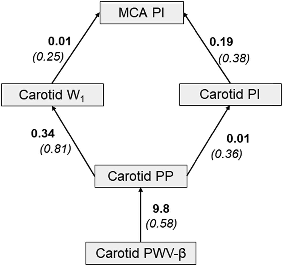
Path analysis highlighting inter-relation between carotid artery stiffness, carotid hemodynamic pulsatility and cerebral hemodynamic pulsatility in children. Presented as unstandardized (standardized) coefficients.
Discussion
Our primary findings support a relationship between extracranial vascular stiffness and intracranial hemodynamic pulsatility in children. Carotid artery stiffness was associated with carotid pressure pulsatility, which in turn was associated with carotid flow pulsatility and ultimately cerebral flow pulsatility. Wave intensity analysis offered insight into the genesis of extracranial hemodynamic pulsatility. Left ventricular ejection into a stiffer/thicker carotid artery may have increased forward compression wave intensity, resulting in enhanced transmission of flow pulsatility into the cerebrovascular bed.
Additional findings of interest should be noted. Carotid pressure and flow pulsatility were both associated with IMT suggesting a relationship between regional pulsatile hemodynamic load and detrimental vascular remodeling in children.7 Carotid AIx was inversely associated with NA suggesting that AIx and NA may capture different aspects of wave reflections (i.e. systemic/global vs regional) in children. Aortic stiffness was not associated with carotid stiffness or carotid/cerebral hemodynamic pulsatility in children. Age and BMI were correlates of cfPWV but not carotid PWV in children and this is consistent with previous observations in adults.8 The aorta and carotid artery “age” at different rates in response to different CVD risk factor exposure.8
Examination of vascular-hemodynamic function in childhood may offer a window into cardiovascular and cerebrovascular risk in adulthood.9 Accumulation of CVD risk factors in childhood influences both arterial stiffness/thickness and cognitive function in young adulthood.10–12 Our findings demonstrate that associations between large artery stiffness and cerebral hemodynamic pulsatility manifest at a young age. Whether targeting arterial stiffness in childhood abrogates subsequent cerebrovascular damage and benefits rates of cognitive decline in adulthood requires further scrutiny.13
In conclusion, similar to what has be previously reported in adults,14 extracranial vascular-hemodynamic pulsatility may influence intracranial hemodynamic pulsatility in children. Future research is required to scrutinize the clinical implications of these associations.
Conflicts of interest statement
This study was funded by a grant from the
Appendix A
Supplementary data
Supplementary data related to this article can be found at
References
Cite this article
TY  - JOUR
AU  - Wesley K. Lefferts
AU  - Jacob P. DeBlois
AU  - Brooks B. Gump
AU  - Kevin S. Heffernan
PY  - 2018
DA  - 2018/05/26
TI  - Carotid artery stiffness and cerebral pulsatility in children
JO  - Artery Research
SP  - 64
EP  - 67
VL  - 22
IS  - C
SN  - 1876-4401
UR  - https://doi.org/10.1016/j.artres.2018.05.002
DO  - 10.1016/j.artres.2018.05.002
ID  - Lefferts2018
ER  -