
YI 1.7 Transmural Quantification of Murine Vascular Smooth Muscle Cell Density Distribution from 3D Microscopy Images
- DOI
- 10.2991/artres.k.201209.007How to use a DOI?
- Keywords
- Anatomy biomechanics adaptation; mechanobiology
- Abstract
Purpose: Investigating the biomechanical role of smooth muscle cells (SMCs) in arteries requires knowledge of their structural distributions. Compared to histology, 3D microscopy offers non-destructive ex vivo imaging under realistic conditions [1]. Robust 3D segmentation of SMCs, however, is challenging. We propose a method for automatic SMC quantification, and assessed its potential using a murine SMC apoptosis model.
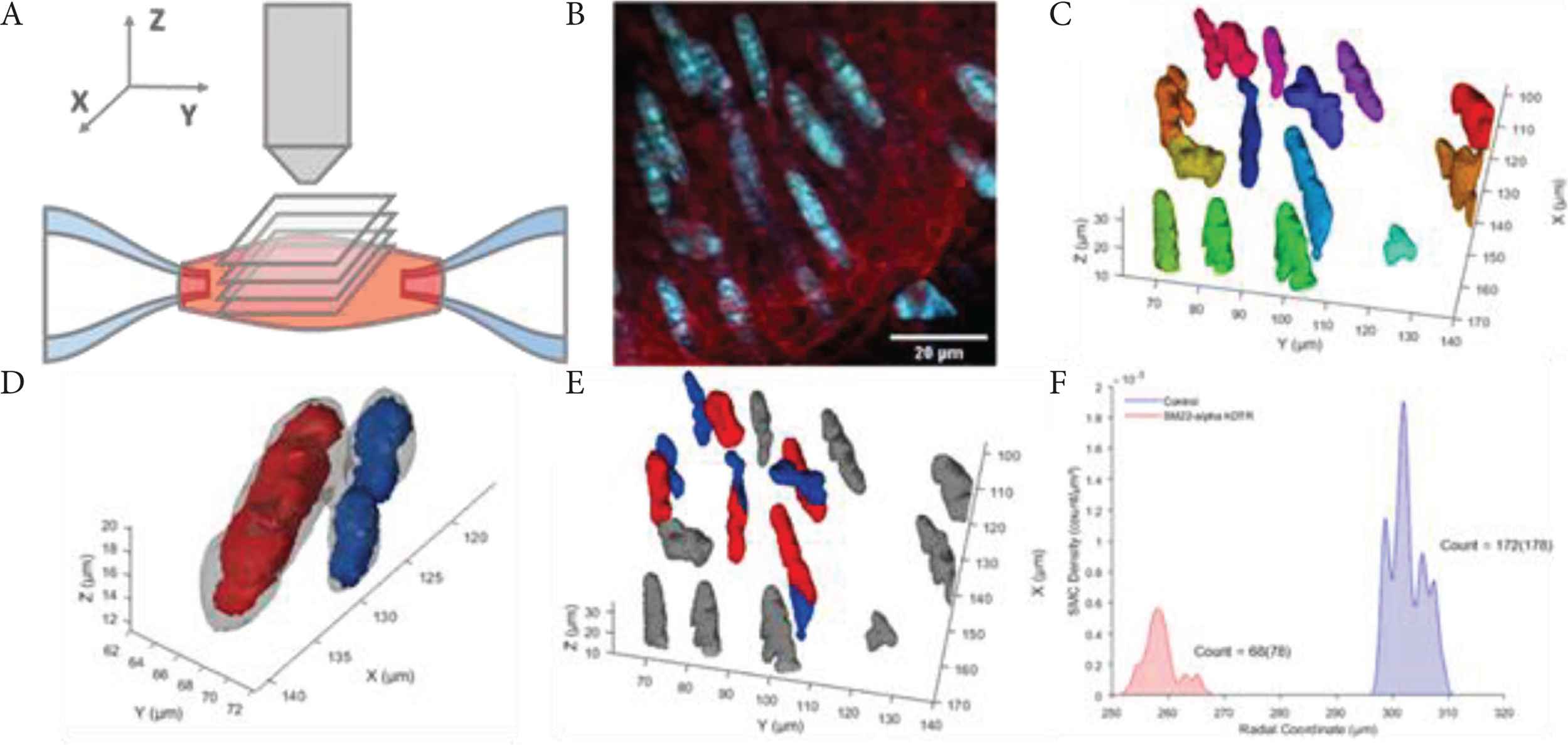
Methods: After euthanasia, carotid arteries (control and with induced SMC apoptosis: SM22α-hDTR [2]) were excised and mounted between micropipettes (Figure A). Nuclei were stained with SYTO41. Arteries were imaged using two-photon microscopy [1], while stretched to in vivo length and pressurised to 100 mmHg (Figure B). Image stacks were processed as follows: 1) deconvolution; 2) nuclei segmentation using vesselness filtering [3,4] (Figure C); 3) cylindrical coordinate system identification; 4) splitting of coincident nuclei, based on cores defined from groups of neighbouring voxels with similar orientations [3] (Figure D and E); 5) cylindrical coordinate system re-identification; and 6) cell density-distribution quantification (Figure F). Segmentation performance was assessed by comparing with manual cell counts.
 Figure
(A) Imaging set-up illustrating acquisition of z-stack of slices. (B) Example slice of 3D stack; cell nuclei are shown in blue while elastin fibres are shown in red. (C) Segmentation results from vesselness filtering of example image stack, colours indicate separated nuclei (step 2, Methods). (D) Coinciding nuclei, corresponding with the left orange nuclei in C, shown in grey, with cell cores shown in red and blue (step 4, Methods). (E) Coinciding nuclei splitting results of nuclei shown in C. Non-split nuclei are shown in grey, while split nuclei are shown in red and blue. (F) Transmural SMC densities and cell counts for one control and one SMC apoptosis sample; manual cell counts are given between parentheses.
Results: Figure E demonstrates the method’s ability to split undersegmented coinciding nuclei. Cell counts were lower in SM22α-hDTR than in control; algorithm-derived counts were comparable to manual (Figure F). The control sample showed multiple SMC layers, while the SM22α-hDTR sample showed a single SMC layer (Figure F), which was confirmed visually.
Conclusion: We developed a precise tool to quantify SMC distributions in ex vivo murine arteries, to facilitate quantitative modelling of SMC biomechanics. We intend to expand the current approach to address cell orientation, shape, and size.
- Copyright
- © 2020 Association for Research into Arterial Structure and Physiology. Publishing services by Atlantis Press International B.V.
- Open Access
- This is an open access article distributed under the CC BY-NC 4.0 license (http://creativecommons.org/licenses/by-nc/4.0/).
REFERENCES
Cite this article
TY  - JOUR
AU  - Koen W.F. van der Laan
AU  - Koen D. Reesink
AU  - Myrthe M. van der Bruggen
AU  - Armand M.G. Jaminon
AU  - Remco T.A. Megens
AU  - Leon J. Schurgers
AU  - Tammo Delhaas
AU  - Bart Spronck
PY  - 2020
DA  - 2020/12/31
TI  - YI 1.7 Transmural Quantification of Murine Vascular Smooth Muscle Cell Density Distribution from 3D Microscopy Images
JO  - Artery Research
SP  - S8
EP  - S9
VL  - 26
IS  - Supplement 1
SN  - 1876-4401
UR  - https://doi.org/10.2991/artres.k.201209.007
DO  - 10.2991/artres.k.201209.007
ID  - vanderLaan2020
ER  -