
P139 AUTOMATIC CLASSIFICATION OF ARTERIAL AND VENULAR TREES IN COLOUR FUNDUS IMAGES
- DOI
- 10.1016/j.artres.2018.10.192How to use a DOI?
- Abstract
Background: Quantitative imaging of retinal arterioles and venules offers unique insights into cardiovascular and microvascular diseases but is laborious. We developed and tested a method to automatically identify Arterial/Venular (A/V) vessels in digital retinal images in conjunction with a semi-automatic segmentation technique.
Methods: Segmentation of blood vessels and the Optic Disc (OD) was performed as previously described [1] using a dataset of X colour fundus images. Using the OD as a reference point a graph representation was constructed using the vessel skeletons. Vessel bifurcations and crossings were identified based on direction and local geometry, and A/V classification was carried out by fuzzy logic classification using colour information. Results were compared with expert classification.
Results: 157 arterial and 150 venular segments were classified. Preliminary Results showed sensitivity, specificity and accuracy of 42.20%, 99.21% and 97.73% for arteries and 50.89%, 98.70% and 97.54% for veins. An example is shown in Figure 1.
Conclusions: Computer-based systems can assess local and global aspects of the retinal microvascular architecture, geometry and topology. Automated A/V classification will facilitate efficient cost-effective assessment of clinical images at scale.
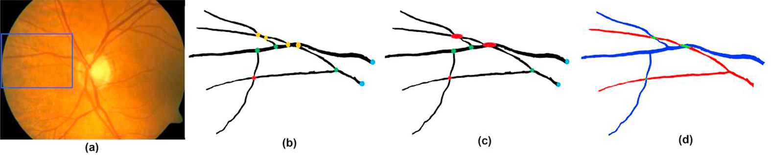 Figure 1
(a) Colour image, rectangle crop area in (b)-(d), (b) segmented blood vessels, red crossing, green bifurcations, blue root and yellow ambiguous points, (c) ambiguous points corrected, and (d) classified vessels, red artery and blue vein.
- Open Access
- This is an open access article distributed under the CC BY-NC license.
Reference
Cite this article
TY  - JOUR
AU  - M Elena Martinez-Perez
AU  - Kim Parker
AU  - Nick Witt
AU  - S.A.McG. Thom
AU  - Alun Hughes
PY  - 2018
DA  - 2018/12/04
TI  - P139 AUTOMATIC CLASSIFICATION OF ARTERIAL AND VENULAR TREES IN COLOUR FUNDUS IMAGES
JO  - Artery Research
SP  - 119
EP  - 119
VL  - 24
IS  - C
SN  - 1876-4401
UR  - https://doi.org/10.1016/j.artres.2018.10.192
DO  - 10.1016/j.artres.2018.10.192
ID  - Martinez-Perez2018
ER  -