
P11.17 CALCIFICATION OF THE THORACIC AORTA ON CHEST X-RAY – ASSOCIATIONS WITH ABDOMINAL AORTIC CALCIFICATION AND PULSE WAVE VELOCITY IN PATIENTS ON RENAL REPLACEMENT THERAPY
- DOI
- 10.1016/j.artres.2011.10.173How to use a DOI?
- Open Access
- This is an open access article distributed under the CC BY-NC license.
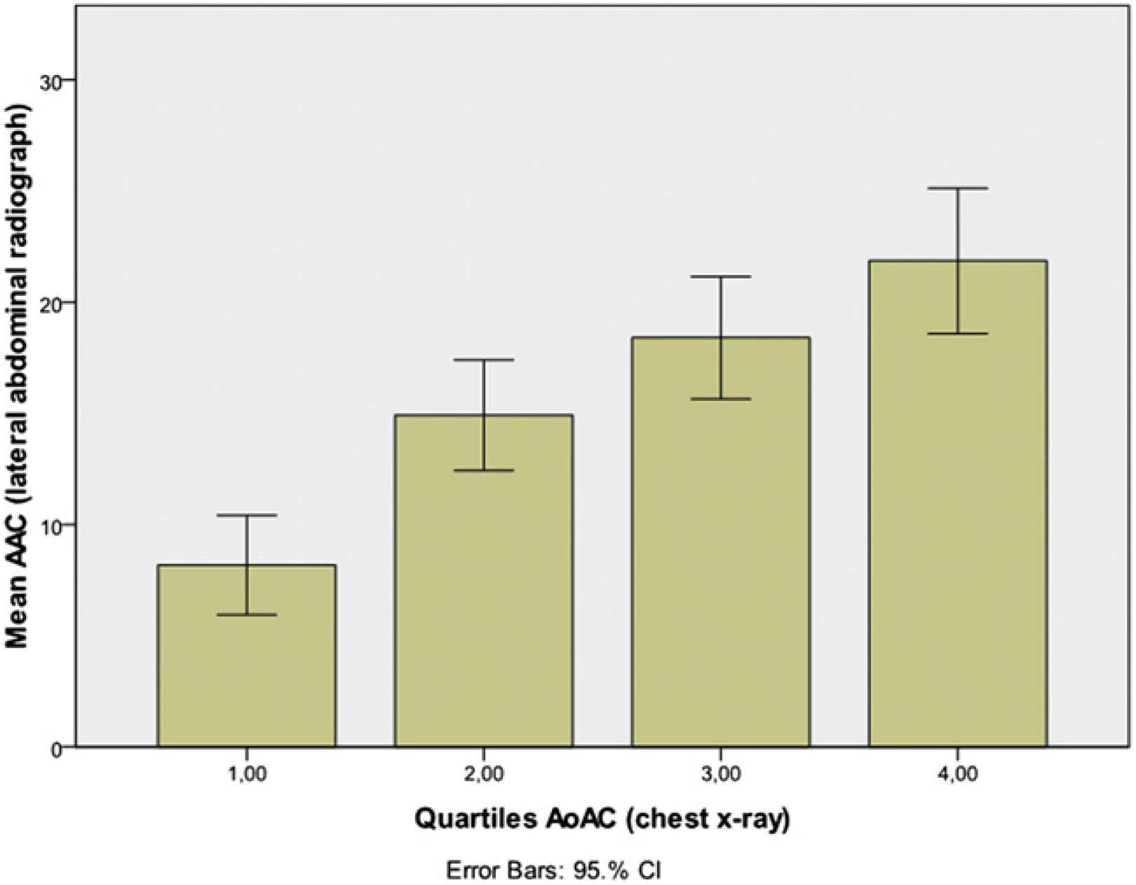
Background: Abdominal aortic calcification (AAC) on lateral abdominal x-ray and carotid-femoral pulse wave velocity (PWV) are independent predictors of mortality and non-fatal CV events in patients on renal replacement therapy (RRT). Guidelines suggest that the presence of AAC can be used to identify patients at high risk. In this study aortic arch calcification (AoAC) on chest x-ray was correlated with the above mentioned established markers of vascular calcification.
Methods: AAC, AoAC and PWV were measured in 88 patients on RRT who were Danish participants in a European multicenter cross-sectional study. Inclusion criteria were: >18 years of age and >3 months of maintenance haemo- or peritoneal dialysis treatment. AoAC was measured using a semi-quantitative method. The cross section of the aortic arch was divided into 16 sectors on a plain frontal chest x-ray. Sectors showing signs of calcified plaques in the form of typically shaped densities were identified. The carotid-femoral PWV was measured using applanation tonometry.
Results: 72% had a AoAC score > 0 compared to 81 % with a AAC score > 0. AoAC was significantly positively correlated with AAC (r=0.69, p<0.001) and PWV (r=0.35, p<0.001). The positive- and negative predictive values of AoAC with respect to AAC were 98% and 32%, respectively.
Conclusion: The presence of calcification on chest x-ray was positively correlated with AAC and PWV. Chest x-ray is inexpensive and frequently obtained in this patient population. We believe it may provide valuable data on calcification and may be used in risk stratification of dialysis patients. Further evaluation is required.
Cite this article
TY  - JOUR
AU  - P. Schjelderup
AU  - J.H. Christensen
AU  - J.D. Jensen
AU  - K.E. Otte
AU  - S. Ladefoged
AU  - P.B. Jensen
PY  - 2011
DA  - 2011/11/29
TI  - P11.17 CALCIFICATION OF THE THORACIC AORTA ON CHEST X-RAY – ASSOCIATIONS WITH ABDOMINAL AORTIC CALCIFICATION AND PULSE WAVE VELOCITY IN PATIENTS ON RENAL REPLACEMENT THERAPY
JO  - Artery Research
SP  - 196
EP  - 197
VL  - 5
IS  - 4
SN  - 1876-4401
UR  - https://doi.org/10.1016/j.artres.2011.10.173
DO  - 10.1016/j.artres.2011.10.173
ID  - Schjelderup2011
ER  -