
Arterial calcification in rheumatoid arthritis
Tel.: +90 242 227 67 12/505 741 15 50; fax: +90 242 227 44 90.
Tel.: +90 242 247 67 21/505 530 51 55; fax: +90 242 227 44 90.
- DOI
- 10.1016/j.artres.2007.10.001How to use a DOI?
- Keywords
- Arterial calcification; Rheumatoid arthritis; Ischaemic necrosis
- Abstract
Arterial calcification most often occurs in patients with end-stage renal disease who undergo hemodialysis or hyperparathyroidism and it is independently associated with cardiovascular events and death. Calciphylaxis is uncommon in patients with rheumatoid arthritis. We present here development of arterial calcification in a patient with rheumatoid arthritis without end-stage renal failure and hyperparathyroidism.
- Copyright
- © 2007 Association for Research into Arterial Structure and Physiology. Published by Elsevier B.V. All rights reserved.
- Open Access
- This is an open access article distributed under the CC BY-NC license.
A 75-year-old woman, who was diagnosed with rheumatoid arthritis before 15 years ago, presented with right foot fingers ulcerations and severe pain. Her examination revealed ischaemic necrosis in the distal parts of the fourth and fifth digits of the right foot and absent dorsalis pedis artery pulse. There was no history of trauma or injury. None of the microorganism grew in swap culture and blood cultures. Bilateral knee radiographs revealed calcified larger vessels (Fig. 1). Amputation of right foot digits was performed. She had been using low-dose steroids (<7.5 mg) and methotrexate for 8 years and had had chronic renal insufficiency for 5 years. Creatinine clearance had remained among 30–45 ml/min and she had never received treatment of hemodialysis or peritoneal dialysis. Levels of parathormon, calcium and phosphorus were normal.
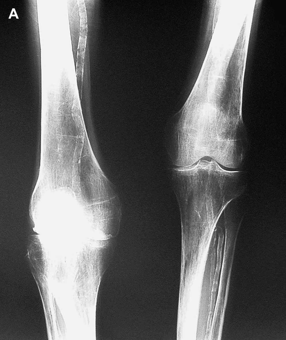
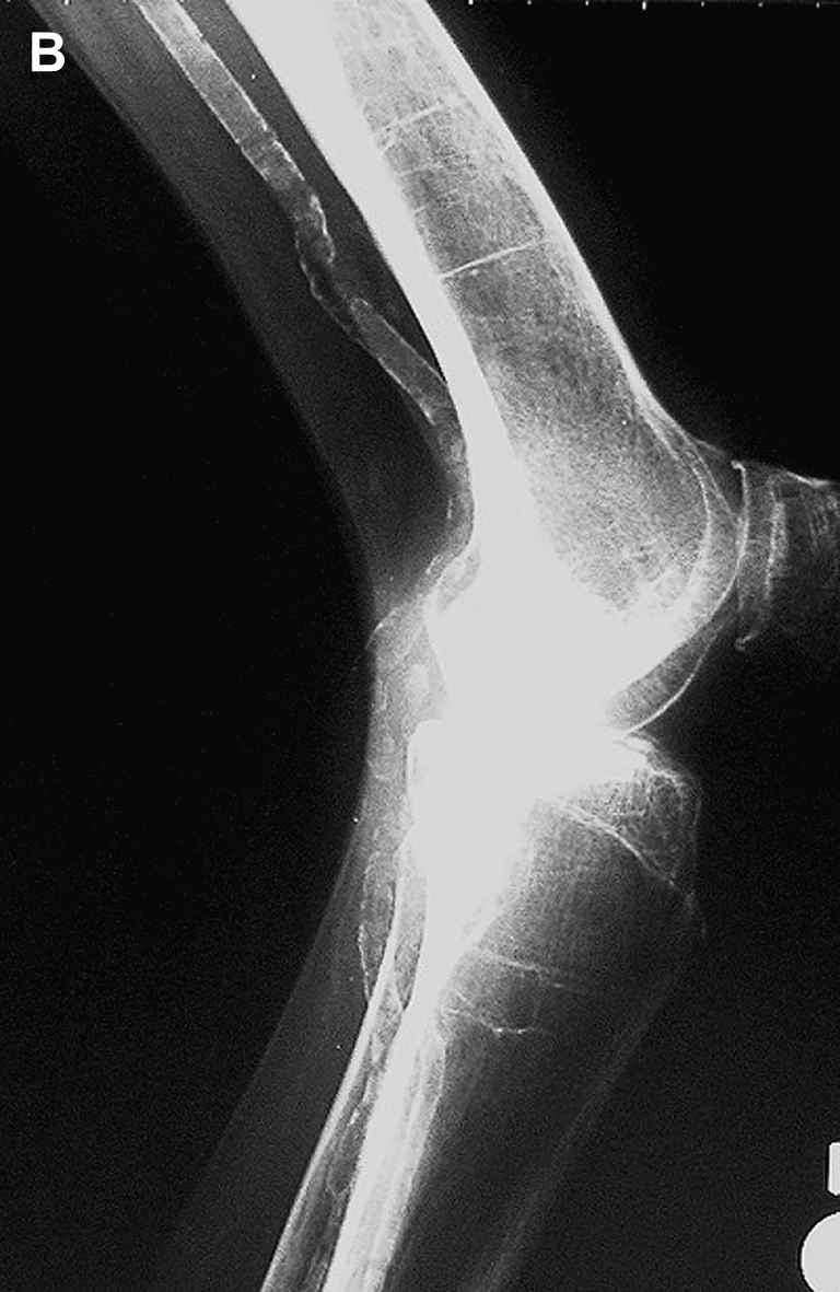
Bilateral knee radiographs show calcified larger vessels and joint destruction in the right knee (anterior and lateral views).
Calcification of vessels most often occurs in patients with end-stage renal disease who undergo hemodialysis and in patients with hyperparathyroidism. Vascular calcification is independently associated with cardiovascular events and death. The prognosis is poor and necrosis occurs at sites of arterial obstruction. This disorder may cause painful skin ulcerations and necrosis at lower limbs and penis. Sometimes amputation of these areas is unavoidable.1 Two rheumatoid arthritis patients with development of calciphylaxis have been reported in the literature up to now.2,3 Treatment with steroids and methotrexate for long periods was proposed to be one of the possible causes of calciphylaxis development.
References
Cite this article
TY  - JOUR
AU  - Veli Yazisiz
AU  - Ali Berkant Avci
AU  - Ender Terzioğlu
PY  - 2008
DA  - 2008/02/21
TI  - Arterial calcification in rheumatoid arthritis
JO  - Artery Research
SP  - 49
EP  - 50
VL  - 2
IS  - 1
SN  - 1876-4401
UR  - https://doi.org/10.1016/j.artres.2007.10.001
DO  - 10.1016/j.artres.2007.10.001
ID  - Yazisiz2008
ER  -