
“Trifurcation” of femoral artery
- DOI
- 10.1016/j.artres.2012.09.004How to use a DOI?
- Keywords
- Femoral artery ramification; Lateral circumflex femoral artery; Thigh flaps
- Abstract
The present study describes analytically a rare case in which a trifurcation of Femoral Artery in Superficial Femoral Artery, Deep Femoral Artery and Lateral Circumflex Femoral Artery was found during dissection of a cadaver. This variation can be considered as a subdivision of Lateral Circumflex Femoral Artery arising from Femoral Artery (frequency: 15% according to Lipper and Pabst) but diameters of the vessels (Lateral Circumflex Femoral Artery was measured at 0.4 cm, of Deep Femoral Artery at 0.55 cm, Femoral Artery at 1 cm and Superficial Femoral Artery at 0.6 cm) and small distance between origin of Deep Femoral Artery and inguinal ligament indicate instead a true trifurcation rather than a simple subdivision. The anatomic variations of the branching pattern of the femoral region arteries concerning both their origin and course are especially important due to the numerous operations performed in the femoral region that implicate various specialties such as orthopedic surgery, plastic surgery, vascular surgery, general surgery and invasive cardiology.
- Copyright
- © 2012 Association for Research into Arterial Structure and Physiology. Published by Elsevier B.V. All rights reserved.
- Open Access
- This is an open access article distributed under the CC BY-NC license.
Brief report
During the dissection of the left femur of a wholly preserved in 10% formalin cadaver in the macroscopic laboratory in Department of Anatomy, Faculty of Medicine, National and Kapodistrian University of Athens the femoral artery seemed to follow an uncommon pattern of ramification.
Common Femoral Artery (CFA) trifurcated into Superficial Femoral Artery (SFA), Deep Femoral Artery (DFA) and Lateral Circumflex Femoral Artery (LCFA). The ramification point was at 1.9 cm from the inguinal ligament. Diameter of LCFA was measured at 0.4 cm, of DFA at 0.55 cm, of the CFA, 1 cm and of SFA at 0.6 cm (Fig. 1). Relative size of the vessels indicates a true trifurcation and not a coincidence of the branching point of DFA and LCFA. No other anatomic variations concerning the femoral region were found on either the right or left femur region of this cadaver.
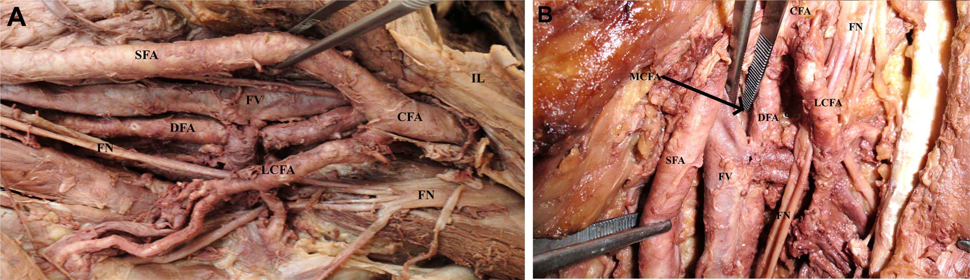
A: Lateral view of femoral region dissection; B: Frontal view of femoral region dissection; FA: Femoral artery; SFA: Superficial femoral artery; DFA: Deep femoral artery; LCFA: Lateral circumflex femoral artery; MCFA: Medial circumflex femoral artery; FV: Femoral vein; FN: Femoral nerve; SN: Saphenous nerve; IL: Inguinal ligament.
Already in 1928 Adachi1 in his classical textbook “Anatomie der Japaner, das Arteriensystem der Japaner” classified ramification patterns of CFA in 8 types and measured their frequency. Lippert and Pabst2 also classified ramification patterns in 8 types. Advances in surgery inspired researchers to propose new classification systems, as the one by Vazquez et al.3 which takes in consideration relative position of Medial Circumflex Femoral Artery (MCFA) and LCFA. Siddharth et al.4 proposed a classification system for LCFA origin and so did Fukuda et al.5 that proposed a classification system focused on LCFA and occurred from its use as arterial graft for Coronary Artery Bypass Grafting (CABG) (Table 1). Our case cannot be precisely classified. It seems to respond to type III of Adachi or Lippert classification, type II of Siddharth classification, intermediate to F1 and F2 of Fukuda classification and somewhat to type IIb of Vazquez classification. Frequency of this variation ranges from 8 to 25%, most researches swarming around 10%, as is the overall frequency.
| Adachi1 classification | Lippert and Pabst2 classification | ||||
|---|---|---|---|---|---|
| I | LCFA and MCFA arising from DFA | 63.2% | I | LCFA and MCFA originating from DFA | 58% |
| II | MCFA arising from SFA | 15% | II | MCFA originating from SFA | 18% |
| III | LCFA arising from SFA | 14.4% | III | LCFA originating from SFA | 15% |
| IV | LCFA and MCFA arising from CFA | 3.6% | IV | LCFA and MCFA originating from CFA | 4% |
| V | MCFA originating from LCFA which originates from SFA | 2.2% | V | As I or II with a branch of LCFA arising from CFA | 3% |
| VI | MCFA and Ld of LCFA from SFA | 0.8% | VI | LCFA and MCFA arising from CFA by a common trunk | 1% |
| VII | MCFA and Ld of LCFA from SFA | 0.5% | VII | Absent MCFA | <1% |
| VIII | Absent MCFA | 0.3% | VIII | DFA originating from external iliac artery | <1% |
| Vazquez3 classification | |||||
| I | Both arteries arise from DFA | 79% | Ia | MCFA origin proximal to LCFA origin | 53.2% |
| Ib | LCFA origin proximal to MCFA origin | 23.4% | |||
| Ic | Both arteries arising from a common trunk | 23.4% | |||
| II | One artery arising from DFA and one from SFA | 20.5% | IIa | MCFA arising from SFA | 77.8% |
| IIb | LCFA arising from SFA | 22.2% | |||
| III | Both arteries arise from SFA | 0.5% | |||
| Siddharth4 classification for LCFA | Fukuda5 classification | ||||
| I | From DFA | 67% | D | LCFA originating from DFA, also a branch of LCFA arising from DFA with LCFA itself | 78.6% |
| II | From SFA | 16% | F1 | LCFA arises from FA above DFA origin | 10.3% |
| III | La from SFA and Ld from DFA | 3% | F2 | LCFA arises from FA below DFA | 5.3% |
| IV | Common origin of DFA, LCFA, MCFA | 5% | F3 | LCFA arises above DFA and a branch from CFA | 3.1% |
| V | Common origin with MCFA | 5% | DF1 | Ld arises from FA and La from DFA | 3.1% |
| VI | Absent LCFA. La and Ld from DFA | 4% | DF2 | The LFCA originates from the DFA and a major branch of the LFCA arises from the FA below the origin of the DFA. | 3.1% |
CFA: Common femoral artery; SFA: Superficial femoral artery; DFA: Deep femoral artery; LCFA: Lateral circumflex femoral artery; MCFA: Medial circumflex femoral artery; Ld: Descending branch of LCFA; La: Ascending branch of LCFA.
CFA ramification patterns.
Another anatomic aspect of significance is correlation of the distance of the branching point of FA from the inguinal ligament with FA ramification pattern. This distance has been measured in a series of studies3 (Siddharth et al.: 44 mm, Dixit et al.: 47.5 mm, Bannister et al.: 35 mm, Snell RS: 40 mm, Vuksanović-Božarić et al.: 37.5 mm, Praksh et al.: 42 mm). There seems to be a correlation between long distance of the branching point and the inguinal ligament and origins of MCFA and LCFA from CFA.3 Small distance measured in this case presentation (19 mm) seems to avoid this “rule” and adds to the particularities of this case.
At 10 mm embryo arterial supply of the leg is provided by sciatic artery (SA) while CFA just starts its development. Various anastomoses develop between those arteries until CFA becomes the main supplier of the lower limb. DFA and its branches seem to be one of these main anastomoses. Anatomic variations of the CFA ramification pattern are attributed to an arrest in development.2
Selective or radial dissection of the femoral region is necessary for a large number of procedures of general surgery, vascular surgery, orthopedics surgery and plastic surgery including femoral hernias, femoral artery obstruction, hip joint surgery and harvesting of various flaps.
Descending branch of LCFA (Ld) is of particular interest for cardiac surgery because it is a vessel that can be used for CABG. LCFA system is important for the creation of pedicled or free flaps such as Tensor Fasciae Latae Perforator Flap (based on ascending branch of LCFA, La), anteromedial thigh perforator flap (based on perforator flaps of Ld) and anterolateral thigh perforator flap (based on medial branch of Ld) used for restoring defects of face, oral cavity, skull base, knee and proximal lower leg.6
Conflicts of interest
None.
Funding source
None.
References
Cite this article
TY  - JOUR
AU  - Theodore Troupis
AU  - Adamantios Michalinos
AU  - Lambros Markos
AU  - Alexandros Samolis
AU  - George Tsakotos
AU  - Dimitrios Dimitroulis
AU  - Dionysios Venieratos
AU  - Panayiotis Skandalakis
PY  - 2012
DA  - 2012/10/02
TI  - “Trifurcation” of femoral artery
JO  - Artery Research
SP  - 106
EP  - 108
VL  - 7
IS  - 2
SN  - 1876-4401
UR  - https://doi.org/10.1016/j.artres.2012.09.004
DO  - 10.1016/j.artres.2012.09.004
ID  - Troupis2012
ER  -