
High origin of radial artery from the axillary artery: Case report
- DOI
- 10.1016/j.artres.2017.01.003How to use a DOI?
- Keywords
- Radial artery; Variation; Anterior interosseous nerve compression
- Abstract
The axillary artery is the main artery supplying the shoulder region and is clinically important. In the present case we report an arterial variation of the left upper extremity of a 65 year-old female cadaver. The radial artery arose from the axillary artery 2.5 cm below the anterior humeral circumflex artery and above the teres major muscle. It first coursed along the medial aspect of the arm, then it passed laterally between the biceps brachii and the brachialis muscle. Reaching the upper part of the elbow, it coursed medial to the brachioradialis muscle. It did not give off any branches in either the arm or in the forearm regions. It contributed to the formation of the superficial palmar arch in the palm of the hand. Further, the brachial artery entered the cubital fossa and divided into ulnar and common interosseous arteries. The right upper extremity had a normal arterial branching pattern.
Being aware of the variations of the axillary artery may help to prevent diagnostic errors and avoid complications during the surgery of the region.
- Copyright
- © 2017 Association for Research into Arterial Structure and Physiology. Published by Elsevier B.V. All rights reserved.
- Open Access
- This is an open access article distributed under the CC BY-NC license.
Introduction
The axillary artery is the continuation of the subclavian artery. The axillary artery lies between the lateral border of the 1st rib and lower border of the teres major muscle. Throughout its course in the axillary region, it is in close relation with the cords and branches of brachial plexus. The artery is customarily divided into three parts according to its relation with the pectoralis minor muscle. Conveniently, each segment gives off a corresponding number of branches. The axillary artery may bifurcate into 2 branches and this is referred to as the “doubling of brachial artery”.1 The axillary artery at the distal border of the teres major muscle continues as the brachial artery. The brachial artery ends about a centimeter distal to the elbow joint, at the level of the neck of the radius. The brachial artery normally bifurcates into the radial and ulnar arteries at the elbow region. Anatomical variations of this artery occur in almost 20% of the cases and are commonly found in routine dissections or in clinical practice.2,3 Six different variations have been identified so far, with the brachial artery following a superficial course (superficial brachial artery) being the most common, whereas the existence of a doubled brachial artery, or a high origin of the radial artery (brachioradial artery) is rarer.1,4 The brachial artery may be absent in rare cases (accessory brachial artery),5 may be divided at a higher level,6 it may trifurcate7,8 or there may be accessory branches that may or may not bifurcate into radial and ulnar arteries.9 Being aware of vascular variations is important for both surgeons and radiologists, and may prevent diagnostic errors.10
Case report
The region of the right arm of a 65 year-old female cadaver was exposed during routine dissection. The axillary artery and its branches were identified. The first two parts of the axillary artery and its branches were normal. The third part of the axillary artery gave rise to the radial artery 2.5 cm below the origin of the anterior humeral circumflex artery and above the teres major (Fig. 1a and b). The radial artery ran along the medial aspect of the arm (Fig. 2a and b) and continued laterally between the biceps brachii and the brachialis muscles (Fig. 1a, b). At the elbow, the radial artery coursed along the medial aspect of the brachioradialis muscle. It gave off no branches either in the arm or in the forearm region. The remaining axillary artery followed its normal anatomical course as the brachial artery to the cubital fossa where it divided into ulnar and common interosseous arteries. The right upper extremity had a normal arterial branching pattern. The radial artery contributed to the formation of the superficial palmar arch in the palm of the hand. The right upper extremity had a normal arterial branching pattern.
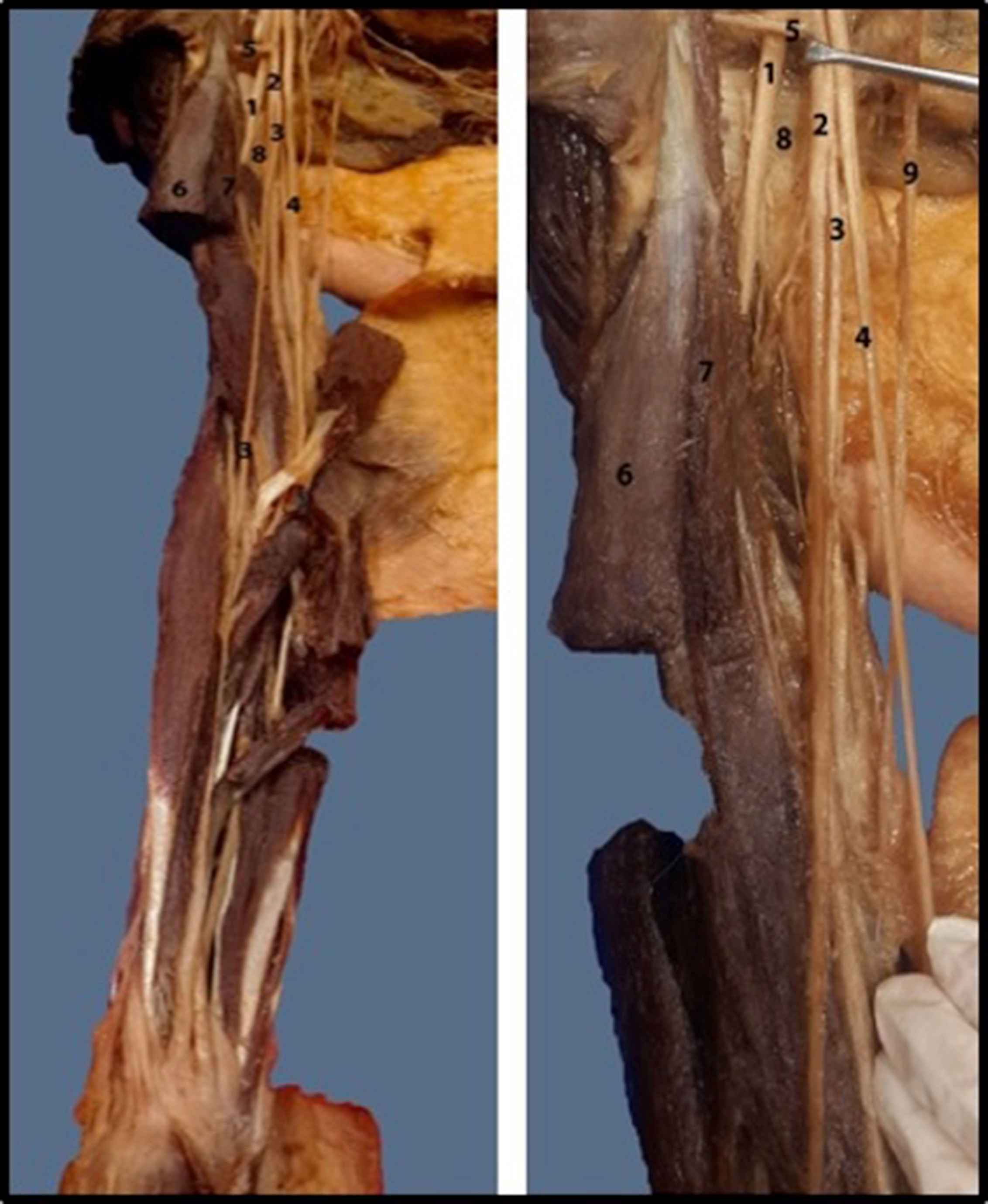
a, b. Cadaveric appearance of the high origin of radial artery from the axillary artery. Abbreviations. 1: Musculocutaneous nerve 2: Axillary nerve 3: Radial artery 4: Median nerve 5: Anterior circumflex humeral artery 6: Biceps brachii muscle 7: Long head of the biceps brachii muscle 8: Teres major 9: Medial cutaneous nerve of arm/forearm.
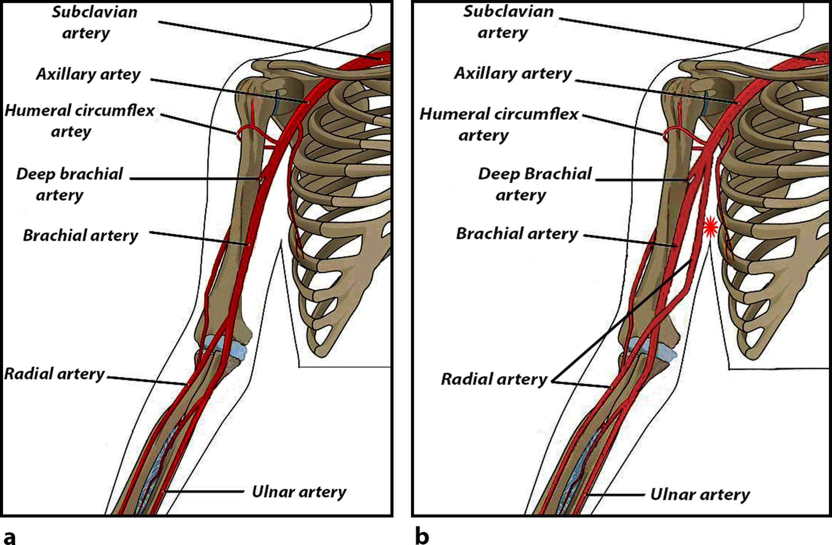
a. Illustration of a normal anatomical course of radial artery. b. Illustration of a variation of the radial artery as being originated from the upper level respectively.  High origin of the radial artery.
Discussion
Variations of the arterial patterns in the human upper extremities have been the subject of many anatomical studies. A review of the literature showed many variations in the axillary artery. In the present case we report a high origin of the radial artery from the axillary artery. During embryogenesis, the vascular plexuses of the limb-buds are initially supplied by four or five consecutive intersegmental branches of the dorsal aortae as the artery passes peripherally at the levels at which the limb-buds are situated. Very early, however, the lateral branches of the seventh cervical and branches of the fifth lumbar intersegmental arteries become much enlarged to form the axial arteries of the upper and lower limbs respectively.
In the arm this axial artery terminates in a capillary plexus from which, later, digital branches arise. The proximal part of the artery can be recognized as the brachial artery, its distal portion forms the interosseous artery. The ulnar and radial arteries arise comparatively late in development as new vessels.11 The arterial anomalies in the upper limb are the defects in the embryonic development of the vascular plexus of the upper limb bud.12 It has been speculated that an arrest at any stage of development may be the cause of regression, or that the retention or reappearance of a vessel may occur and this may lead to variations in the arterial origins and courses of the major upper limb vessels.5
The vascular variations of the upper limb are fairly common and have been reported extensively. The axillary artery is the second most susceptible artery to lacerations caused by violence and thus knowing the possible variations of the branching pattern of the vessels is important in the field of reparative surgery as well as during angiography procedures.13 Williams14 presented a case where the axillary artery ruptured in an attempt to reduce old a dislocations, which is particularly likely when the artery is adherent to the articular capsule.
Branches of the upper limb arteries have been used for coronary bypasses and as flaps in reconstructive surgery. Knowledge of normal and variant arterial arrangement of the human upper limb may serve as a useful guide for both reparative surgeons and radiologists.10
In an attempt to get correct specific diagnostic images for vascular lesions with contrast arteriography and duplex ultrasonography, examinations of images of the vascular patterns of the axillary may lead to misinterpretation of the data where an unusual or complicated variation of the artery exists.15 Therefore, the knowledge of variations of the axillary artery and evaluation of its possible route is very important for the prevention of diagnostic errors and avoid complications for surgeons working in the axillary region.
Conflict of interest statement
The authors declare no conflict of interest to disclose.
References
Cite this article
TY  - JOUR
AU  - Merve Özgür
AU  - Murat Gölpınar
AU  - Yasin Arifoğlu
AU  - Safiye Çavdar
PY  - 2017
DA  - 2017/02/09
TI  - High origin of radial artery from the axillary artery: Case report
JO  - Artery Research
SP  - 39
EP  - 42
VL  - 17
IS  - C
SN  - 1876-4401
UR  - https://doi.org/10.1016/j.artres.2017.01.003
DO  - 10.1016/j.artres.2017.01.003
ID  - Özgür2017
ER  -