
Combinatorial effect of lower extremity blood flow restriction and low intensity endurance exercise on aorta of old male rats: Histomorphological and molecular approach
- DOI
- 10.1016/j.artres.2018.10.226How to use a DOI?
- Keywords
- Blood flow restriction; Exercise training; Apelin receptor; Angiotensin receptor; Aorta wall thickness
- Abstract
Objective: Given the importance of knowing the effects of blood flow restriction (BFR) plus exercise training (BFR training) on the cardiovascular system, present study conducted to determine the effect of combination of BFR with low intensity endurance exercise on aorta of old rats.
Methods: Animal groups were control (CTL), sham (Sh), BFR, exercise (Ex), sham + exercise (Sh + Ex), and BFR + exercise (BFR + Ex). BFR induced by partial closing of the bilateral femoral arteries and duration of exercise was 10 weeks.
Results: BFR induced increasing of aorta wall thickness and decreasing its elastin/collagen ratio. Training alone increased the expression of apelin (P < 0.05) and angiotensin AT2 receptor (AT2R) (P < 0.01 versus CTL, Sh and P < 0.05 versus BFR groups) and the elastin/collagen ratio (P < 0.001) but, decreased the expression of angiotensin AT1 receptor (AT1R) (P < 0.01 versus CTL, Sh and BFR groups), the AT1R/AT2R ratio and the wall thickness (P < 0.001) of aorta. In Ex + BFR group, the expression of mentioned receptors increased and reached to more than levels of Ex groups, and the AT1R/AT2R ratio reduced in comparison with CTL, Sh and BFR and was higher than training groups (P < 0.5). Exercise also reversed the negative effect of BFR on elastin/collagen ratio and wall thickness.
Conclusion: The beneficial effects of low intensity endurance exercise on aorta of aged male rats are conserved partially in presence of BFR.
- Copyright
- © 2018 Association for Research into Arterial Structure and Physiology. Published by Elsevier B.V. All rights reserved.
- Open Access
- This is an open access article distributed under the CC BY-NC license.
Introduction
The prevalence of cardiovascular diseases, the most common cause of death, increases dramatically with age.1 Age-dependent remodeling of the aorta and arterial branches including hypertrophy, collagen accumulation and endothelium abnormalities, are considered as key factors in hypertension which increases the chance of mortality.2,3 Renin-Angiotensin system plays an important role in cardiovascular homeostasis and in the development of cardiovascular diseases.4,5 Angiotensin II has two type receptors, AT1R and AT2R. The biological and primary effects of angiotensin II through AT1R are including smooth muscle contraction, cell proliferation, cell migration, extracellular matrix aggregation, and inflammation.6–8 Activating the AT2R increases the production of NO and inhibits norepinephrine secretion and thus regulates blood pressure and inhibits hypertrophy and fibrosis.9,10 On the other hand, the apelin receptor (APJ) is also present in the heart, coronary arteries and aorta.11 Apelin has potent vasodilatory, blood pressure reducing and cardiac inotropic effects.12–17 It has two-dimensional role in vascular tonicity which includes an endothelium-dependent action to produce NO and dilatation of the vessel and a non-endothelium-dependent effect, which can cause a contraction of the vessel in the absence of the endothelium.18 Apelin 13 bonds to APJ receptors in vascular endothelial cells and activates NO synthesis.19,20 There is a reverse arrangement between the apelin system and its receptor (APJ) with the angiotensin system.21 While apelin is a vasodilator and reduces blood pressure, angiotensin II increases vascular tonicity.22
Aging is associated with an increase in expression of the type I angiotensin II receptors (AT1Rs),23 and a decrease in expression of the APJ receptors.24 Moderate aerobic exercise reduces the age-dependent effects of angiotensin II, which in turn reduces the remodeling changes and improves endothelial function of the aorta in hypertensive rats.25 It also reported that long term swimming exercise reverses the down-regulation of the cardiovascular apelin/APJ system induced by hypertension in rats and effectively reduces hypertension and pathological cardiac hypertrophy.26 In addition, sports activities improve the function of vascular endothelium and prevent atherosclerosis.26–28 Blood flow restriction (BFR) with low-intensity exercise (Kaatsu model) is used as a way to increase the strength and hypertrophy of skeletal muscle in various exercises, such as resistance exercises, hiking, and cycling. This type of sport is popular in Japan and has recently become an interesting research topic.29 In this method, a pressure cuff is typically used to close the proximal part of the upper or lower limbs30–32 so that, blood flow to the muscle in the resting state is well done, but during the activity, the limb becomes ischemic. This method apparently is suitable for the aging people, who are not able to carry out heavy exercises due to some degrees of joint damage and muscle weakness or with cardiovascular disease. In addition to inducing changes in muscle volume and strength, Kaatsu method may have hemodynamic and cardiovascular effects.33 However, so far, the cardiovascular effects of this method in senescence have been less addressed and there is a concern regarding the potentially risk of deleterious cardiovascular events especially in aging individuals performing resistance training exercise with BFR. Recently, we showed that mild endurance exercise plus BFR altered the expression of angiotensin II and apelin receptors of heart which was associated with cardiac hypertrophy and improved the ventricular conductivity of the aging rats.34 Regarding the role of renin-angiotensin and apelinergic systems in structural changes of the aorta during the aging process, the purpose of this study was to answer the question whether combination of limb blood flow restriction and low-intensity endurance exercise has any effect on histological indices and angiotensin II and apelin receptors of aorta in old male rats?
Materials and methods
Animals
Experiments were performed on 60 male rats (24–22 months) weighing 450–350 g. The animals randomly were divided into 6 equal groups and were kept under conditions of 12 hours of darkness and 12 hours of light and free access to water and food.
Ethical approval
All experimental methods were approved by the Ethics Committee of Kerman University of Medical Sciences, Kerman, Iran (Permission No. IR.KMU.RE.1395/275).
Study design
Animal groups were control (CTL), sham (Sh), Blood flow restriction (BFR), Exercise (Ex), Sham + exercise (Sh + Ex) and Blood flow restriction + Exercise (BFR + Ex) groups. CTL group was not subjected to any intervention and they were taken care of for 11 weeks. Sham (Sh), animals group were anesthetized by ketamine (100 mg/kg) and xylazin (10 mg/kg)35 and the inner surfaces of groins of this group were incised to access femoral arteries. After separation of femoral arteries from femoral sheaths, a small amount of powder of penicillin was shed on the wounds. Then the sites of surgery were sutured and animals were under observation for 11 weeks. Blood flow restriction (BFR) group was operated similar to sham group. Then a 0.014 inch diameter steel wire was placed on the femoral artery and it was tightly fixed using a silk stitch thread (4-0). Then the wire removed and incision sites were stitched. In this condition during contraction and training but not resting, the muscle is experiencing ischemia.34–36
In Ex group, the intact animals were monitored for one week after division and then trained with low intensity exercise for 10 weeks according to the method explained in the previous studies.34,37 Animals of Sh + Ex group were operated as explained for sham group and after one week recovery period, they subjected to low intensity treadmill exercise for 10 weeks.
BFR + Exgroup was subjected to surgery as BFR group and also trained with low intensity exercise for 10 weeks after recovery period.34,35,37
Forty-eight hours after the last training session, all animals groups were sacrificed under deep anesthesia using thiopental sodium (100 mg/kg). Then the thorax and abdomen was opened and descending aorta was removed to evaluate the histological parameters and to measure the values of the APJ, AT1R and AT2R proteins.
Training protocol
Exercise groups were exercising 5 days/week for 10 weeks as previously reported.34,35,37 In brief: During the familiarization phase (the first week), 15 minutes waking on a rodent treadmill with speed of 7.5 m/min was considered. During overload phase (day 8–63) the treadmill speed progressively increased to 15 meters/m and the duration of the exercise sessions gradually increased to 60 minutes. At the last week, the treadmill speed and duration of exercise sessions were maintained as 15 meters/m and 60 minutes respectively. The treadmill angle was 0° during protocol. It should be noted that the first 5 minutes for heating, and the last 5 minutes to cool down at a speed of 7 meters per minute had considered. In addition, for the purpose of familiarizing the control, sham and BFR groups were placed on the treadmill at least twice a week.34,35,37
Western blot analysis
The western blot method was used to measure the protein levels of angiotensin receptors 1 and 2 and APJ protein. After collecting, the samples were washed in PBS solution. Homogenization of 80 mg of aorta was performed in ice cold RIPA buffer solution (Sigma Aldrich Company, USA; R0278) plus protease inhibitor (Sigma Aldrich Company, USA; S8820). All stages of homogeneity were performed on ice and maintaining the cold conditions by Sony Kid device. In the next stage, the homogenized tissues were centrifuged at 4 °C at 13000 rpm for 20 minutes and the supernatant was isolated and the total protein concentration of aortic samples were measured by Lowry method and Bovine serum albumin was used as standard. After matching the concentrations, 40 micrograms of protein from each sample were electrophoresed on 11% polyacrylamide gels for 75 minutes. After electrophoresis, the proteins separated in the gel were transferred after 100 minutes to PVDF paper and then incubated overnight at 4 °C in a solution of 2% non-fat dry milk (Sigma–Aldrich, St. Louis, MO, USA). Thereafter, After 4 times washing for 5 minutes with TBST buffer the membranes were incubated for three hours with primary antibody against AT1 (1:200; Sc-579; Santa Cruz Biotechnology, USA), AT2 (1:200; Sc-9040; Santa Cruz Biotechnology, USA), APJ (1:200; Sc-33823; Santa Cruz Biotechnology, USA) or GAPDH (1:1000; Sc-47724; Santa Cruz Biotechnology, USA). After 4 times washing for 5 minutes with TBST buffer (Tris-buffered saline, 0.1% Tween 20), the membranes were incubated for one and half hour with horseradish peroxidase-conjugated secondary antibodies include goat anti-rabbit IgG-HRP (1:10,000; Sc-2004; Santa Cruz Biotechnology, USA) for AT1,AT2, APJ and goat anti-mouse IgG-HRP (1:5000; Sc-2005; Santa Cruz Biotechnology, USA) for GAPDH. Then, four times washing was performed for 5 minutes with TBST buffer, and the membrane was incubated for one minute with chemiluminescence substrate (ECL) (Amersham ECL Prime Western Blotting Detection Reagent). In the next step, immune detection was recorded using Chemi Doc XRS + imaging system (Bio-Rad Company, USA) and analyzed by the Image Lab 3 software. The amount of protein expression was normalized by dividing the each receptor band density to the corresponding GAPDH band density.34,38
Histological assessment
The rats were killed and a part of descending aorta of each animal was fixed in 10% neutral buffered formalin for 24 h and after routine tissue processing, the specimen embedded in paraffin. Serial 5 μm thick sections were stained with hematoxylin eosin (H&E) (general morphology), Orcein (elastic fibers detection), and Masson’s trichrome (collagen fibers detection) methods. Stained sections were examined and photographed using an Olympus digital camera attached to Nikon 50i microscope. The thickness of aortic wall was estimated from the average thickness at 4 points of the cross-sectional aortic wall in the H&E-stained sections. Image J software (NIH, Bethesda, MD, USA) was used to measure of elastin and collagen areas. Elastin content was calculated by dividing the elastin-positive area by the cross-sectional media area of the aortic wall, which was measured using the stained sections to highlight the internal elastic lamina and external elastic lamina. Collagen content was calculated by measuring the adventitia layer thickness (collagen-positive area by the cross-sectional aortic wall area). Then the ratio of elastin to collagen was calculated.
Statistical methods
Results are expressed as mean ± SEM. Statistical computing was used SPSS 22 software. The normal distribution of data was evaluated by Shapiro test and statistical analysis performed by using of one way ANOVA and Post hoc Tukey tests. P < 0.05 was taken as statistically significant.
Results
Receptors expression
Low intensity endurance exercise significantly increased the expression of APJ receptor in the rats’ aorta of Ex and Sh + Ex groups in comparison to the CTL, Sh and BFR groups (P < 0.05). Combination of low intensity endurance training with limbs blood flow restriction induced a significant increase in expression of APJ receptor in aorta of rats (P < 0.001 versusCTL, Sh and BFR groups and (P < 0.05 versus Ex and Sh + Ex groups) (Fig. 1).
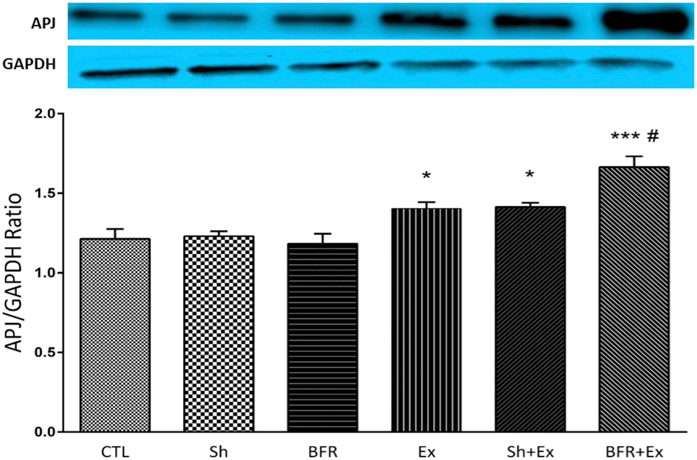
Western blotting results of apelin receptor (APJ) protein expression in various experimental groups. Values are presented as mean ± S.E. The number of animals in each group was 6–8. *p < 0.05 versus CTL, Sham and BFR groups; ***P < 0.001 versus CTL, Sham and BFR groups; # (p < 0.05) versus Ex and Sh + Ex groups. CTL: control group, Sh: sham group, BFR: Blood flow restriction group, Ex: Exercise group, Sh + Ex: Sh + exercise group, BFR + Ex: Blood flow restriction + exercise group.
On the other hand, the value of type 1 angiotensin receptor (ATR1) protein in the aorta of rats showed a significant decrease after 10 weeks of exercise in Ex and Sh + Ex groups compared to CTL, Sh and BFR groups (P < 0.01). When animals subjected to endurance exercise along with limitation of lower limbs blood flow, the effect of exercise on ATR1 protein was masked, so that, this variable in BFR + Ex group did not show any significant difference with CTL, Sh and BFR groups, but increased significantly in comparison with the Ex and Sh + Ex groups (P < 0.001) (Fig. 2). In addition, the value of ATR2 protein significantly increased in Ex and Sh + Ex groups when compared to CTL, Sh (P < 0.01) and BFR (P < 0.05) groups. This protein also was significantly higher in BFR + Ex group (P < 0.001 versus CTL and Sh, and P < 0.01 versus BFR, Ex and Ex + Sh groups) (Fig. 3). Accordingly, the ATR1/ATR2 proteins ratio significantly was lower in Ex and Sh + Ex groups (P < 0.001) and BFR + Ex group (P < 0.05) versus the values in CTL and Sham groups (Fig. 4).
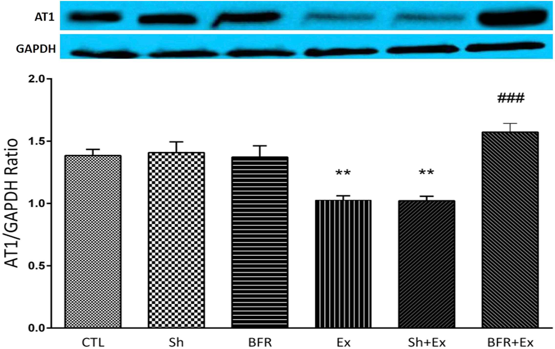
Western blotting results of aortic angiotensin II receptor1 (AT1R) protein expression in various experimental groups. Values are presented as mean ± S.E. The number of animals in each group was 6–8. **p < 0.01 versus CTL, Sham and BFR groups; ###P < 0.001 versus Ex and Sh + Ex groups. CTL: control group, Sh: sham group, BFR: Blood flow restriction group, Ex: Exercise group, Sh + Ex: Sham + exercise group, BFR + Ex: Blood flow restriction + exercise group.

Western blotting results of aortic angiotensin II receptor2 (AT2R) protein expression in various experimental groups. Values are presented as mean ± S.E. The number of animals in each group was 6–8. **p < 0.01 versus CTL and Sh groups; ***P < 0.001 versus CTL and Sh groups; #P < 0.05 versus BFR groups; ##P < 0.01 versus BFR groups. CTL: control group, Sh: sham group, BFR: Blood flow restriction group, Ex: Exercise group, Sh + Ex: Sham + exercise group, BFR + Ex: Blood flow restriction + exercise group.
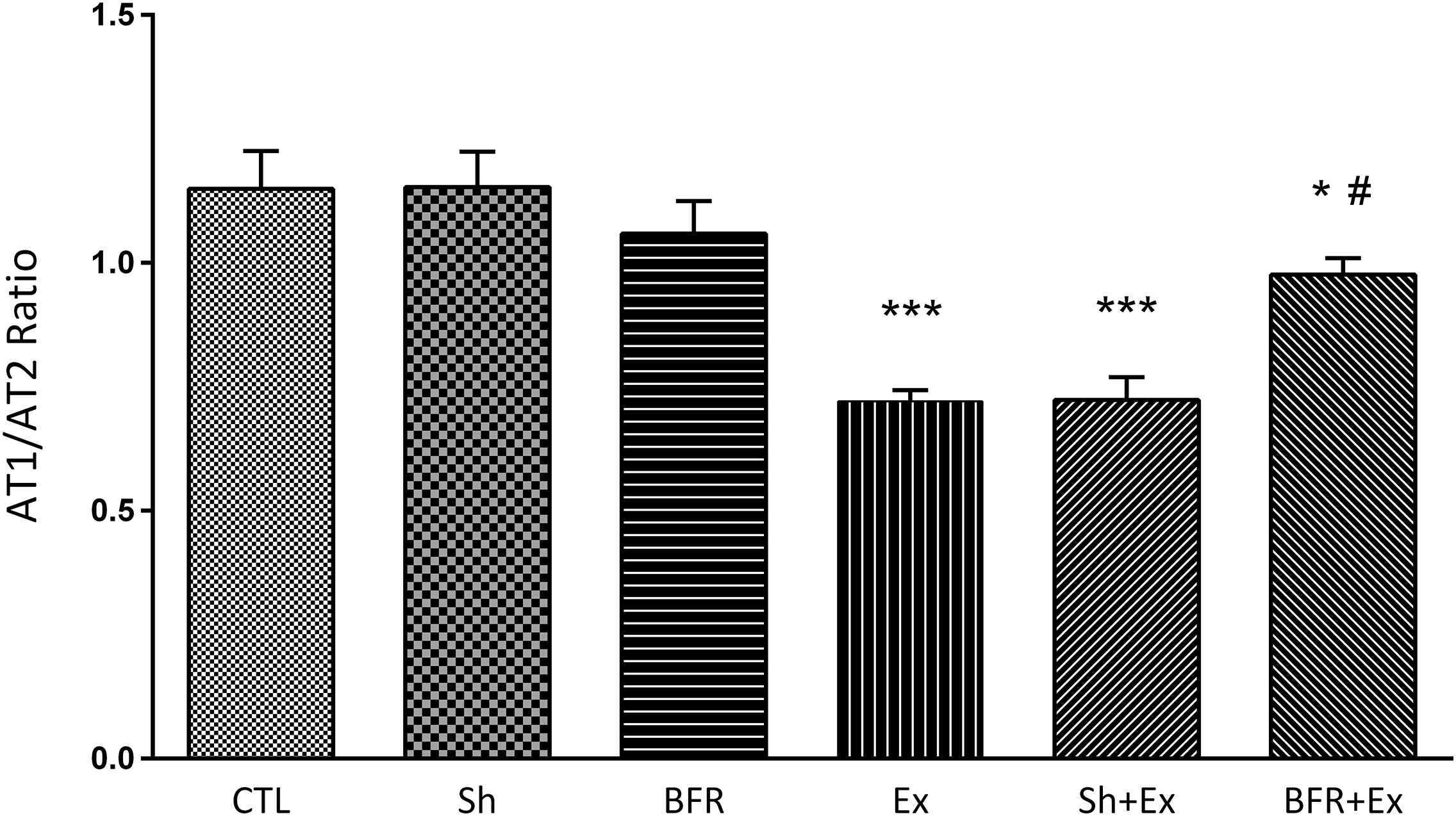
The ratio of aortic angiotensin II receptor1 (AT1R)/angiotensin II receptor2 (AT2R) protein in various experimental groups. Values are presented as mean ± S.E. The number of animals in each group was 6–8. *p < 0.05 versus CTL and Sh groups; ***P < 0.001 versus CTL, Sh and BFR groups; #P < 0.05 versus BFR group. CTL: control group, Sh: sham group, BFR: Blood flow restriction group, Ex: Exercise group, Sh + Ex: Sham + exercise group, BFR + Ex: Blood flow restriction + exercise group.
Histomorpholology
Histological findings showed limbs blood flow restriction alone was associated with reduction in the elastin/collagen ratio when compared with the control and sham groups (P < 0.05). On the other hand, in the exercise groups (Ex and Sh + Ex) this ratio significantly increased in comparison with CTL, Sh and BFR groups (P < 0.001).
In the BFR + Ex group, this ratio was higher than CTL, Sham and BFR groups (P < 0.001), but it was lower than Ex and Sh + Ex groups (P < 0.05) (Fig. 5).
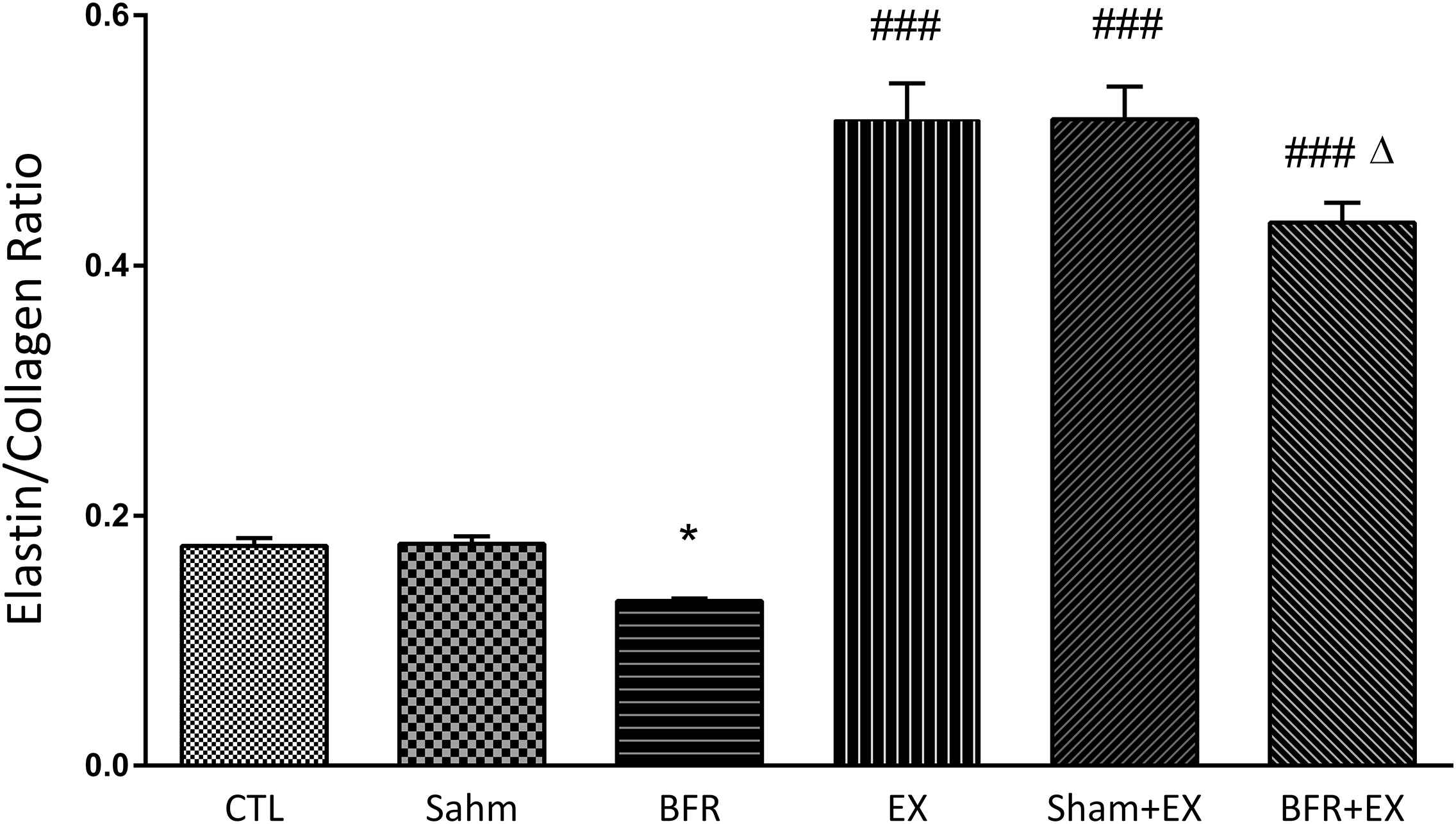
The elastin-collagen ratio of descending aorta in various experimental groups. Values are presented as mean ± S.E. The number of animals in each group was 6–8. *p < 0.05 versus CTL and Sham groups; ###P < 0.001 versus CTL, Sh and BFR groups; Δ P < 0.05 versus Ex and Sh + Ex groups. CTL: control group, Sh: sham group, BFR: Blood flow restriction group, Ex: Exercise group, Sh + Ex: Sham + exercise group, BFR + Ex: Blood flow restriction + exercise group.
Limitation of the bilateral lower extremity blood flow significantly increased the intima-media and total wall thickness of aorta (p < 0.001 versus CTL and Sh groups). These parameters significantly decreased in presence of low intensity endurance exercise in Ex and Sh + Ex groups (p < 0.001 versus CTL, Sh and BFR groups) and also in the BFR + Ex group (p < 0.001 versus CTL, Sh and BFR groups). However, These indices were higher in BFR + Ex than Ex and Sh + Ex groups (P < 0.05) (Figs. 6 and 7).
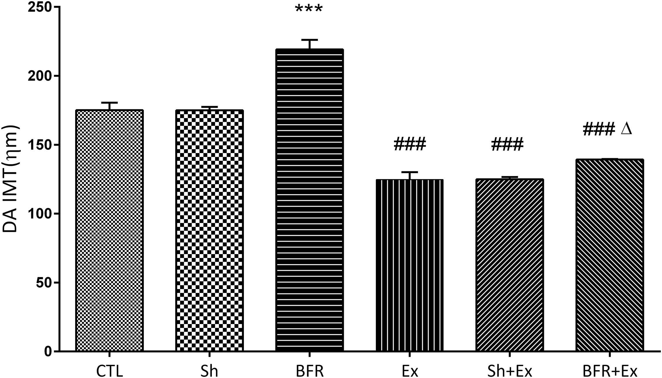
The intima-media thickness of descending aorta in various experimental groups. Values are presented as mean ± S.E. The number of animals in each group was 6–8. ***p < 0.001 versus CTL and Sh groups; ###P < 0.001 versus CTL, Sham and BFR groups; Δ P < 0.05 versus Ex and Sham + Ex groups. CTL: control group, Sh: sham group, BFR: Blood flow restriction group, Ex: Exercise group, Sh + Ex: Sham + exercise group, BFR + Ex: Blood flow restriction + exercise group.
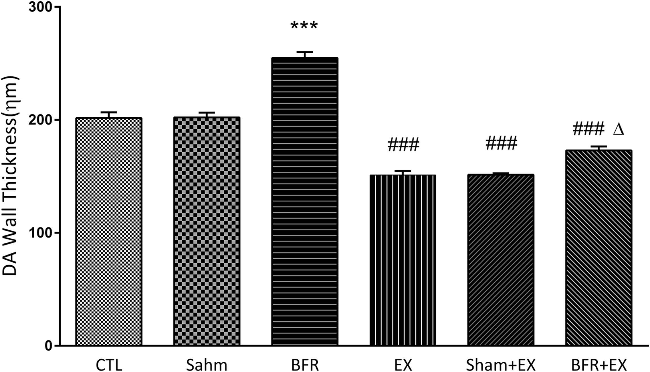
The Wall thickness of descending aorta in various experimental groups. Values are presented as mean ± S.E. The number of animals in each group was 6–8. ***p < 0.001 versus CTL and Sh groups; ###P < 0.001 versus CTL, Sh and BFR groups; Δ P < 0.05 versus Ex and Sh + Ex groups. CTL: control group, Sh: sham group, BFR: Blood flow restriction group, Ex: Exercise group, Sh + Ex: Sham + exercise group, BFR + Ex: Blood flow restriction + exercise group.
Discussion
This study was conducted to investigate the effects of 10 weeks of low intensity endurance exercise along with limited limb blood flow on histomorphological parameters and expression of angiotensin and apelin receptors in aorta of old male rats.
The results showed that low intensity exercise alone significantly decreased the expression of ATR1 but, increased the expression of ATR2 and also apelin receptors. When exercise combined with lower limbs blood flow restriction, the expression of all three mentioned receptors was enhanced and reached to more than levels which observed in exercise alone. Interestingly, the ratio of ATR1/ATR2 was decreased in exercise condition with/without blood flow restriction. In addition, blood flow restriction alone increased the total and intima-media wall thickness of aorta and decreased its elastin/collagen ratio, however combination of this intervention with exercise reversed these effects and improved the elastin/collagen ratio and attenuated the wall thickness of aorta.
In agreement with present study, the positive role of exercise on expression of apelin/APJ and promotion of apelinergic system has been reported. Zhang et al. showed long term swimming exercise increased both apelin and its receptor on aorta and heart of spontaneous hypertensive rats and they suggested that the improving effect of exercise training on hypertension could be mediated by up-regulating the cardiovascular apelin/APJ system.39 In our previous study low intensity exercise increased the apelin receptor in hearts of aging rats and combination of exercise with BFR promoted the effect of exercise34 which is consistent with the finding of present study. Also, in agree with our findings there are evidences that exercise induces down-regulation of ATR1 in heart of aging rats.34,40 In addition exercise improves the expression of ATR2 and attenuates ATR1 and the NADPH oxidase activity in aorta and coronary vessels which resulting in attenuation of AngII-induced vasoconstriction.41,42 As mentioned above, in present study, in exercise plus blood flow restriction, the value of ATR1, ATR2 and ATR1/ATR2 were more than Ex groups, however the ratio of ATR1/ATR2 in this group was less than CTL and sham groups. In present study, molecular findings were consistent with the histological findings, so that the ratio of elastin/collagen significantly increased and intima-media and total wall thickness of aorta reduced in the presence of exercise alone. Despite the fact that BFR alone showed a negative effect on elastin/collagen ratio and increased the intima-media and total wall thickness of aorta, however, these negative effects were masked remarkably by exercise, so that the histological indices in Ex + BFR group were significantly less than CTL, Sh and BFR groups.
Aging is associated with increasing ATR1 receptor,43–45 decreasing ATR245 and APJ receptors46 in the cardiovascular system and also increasing collagen and reducing elastin production in the vessels. Increasing collagen and reducing elastin production during aging leads to increasing of aortic wall thickness and pathological remodeling which can result to hypertension. Stimulation of the AT1 receptor activates fibroblasts and increases the production of collagen levels and decreases elastin, but stimulation of AT247 and APJ receptors48,49 alleviates the effects of AT1 receptors.
It seems in aging rats low intensity exercise alone has beneficial effect on aortic remodeling and corrects the intima-media and total wall thickness by improving the ratio of elastin/collagen partly through modulation of apelin/APJ system and rennin-angiotensin system including ATR receptors balance and relevant molecular signaling cascades. Apparently, blood flow restriction through increasing peripheral resistance can accelerate the aortic remodeling and wall thickening in aging rats. In addition, partial femoral stenosis may change the pattern of blood flow in throat of stenosis and especially immediately after it, from laminar to turbulent. This new flow pattern in turn can be accompanied by change in wall stress and pressure drop in and after the stenosis. These alterations in flow, pressure, and wall stress may cause the release of vasoactive mediators50 from downstream vessels that may modulate the effects of blood flow restriction on aortic remodeling. However, regardless of how BFR induces aortic remodeling effect, it almost completely disappears in the presence of the low intensity exercise training. In consistent of the results of this study, recently we showed that BFR training improve cardiac performance of old rats without significant effect on blood pressure.51 A human study reported that in elderly subjects when low endurance exercise is combined with BFR, it can improves skeletal muscles strength and cross-sectional area and also carotid arterial compliance52 which confirms our findings. It is comes from this fact that the endurance exercise induces increasing of elastin/collagen ratio, which observed in our study, and thereby improves the arterial distensibility and compliance. Of course, it should be remember that reductions in blood flow of muscles especially during resistance exercise activates the exercise pressor reflex (EPR) which considerably contributes to the autonomic cardiovascular response to exercise for increasing the blood pressure through enhancing both cardiac output and peripheral vascular resistance.53 Therefore, EPR activating and its likely contribution to the BFR-mediated cardiovascular response to exercise should be considered in patient with cardiovascular diseases.
In conclusion, findings suggest that blood flow restriction alone may be associated with undesirable aortic remodeling. On the other hand low intensity endurance exercise induced the beneficial physiological remodeling effect on aorta of aged male rats. A part of this beneficial effect is due to increasing of the aortic APJ and AT2 receptors and modulation of ATR1/ATR2 ratio. In presence of both low intensity endurance exercise and BFR, the beneficial effect of exercise on aortic structure is conserved partially and hence increased aortic distensibility and compliance by improving elastin/collagen ratio in aging rats. Further studies are needed to understanding the all of involving mechanisms and generalizing these findings to human.
Abbreviations
- ACE
Angiotensin converting enzyme
- ACE2
Angiotensin converting enzyme type 2
- Ang (1–7)
Angiotensin (1–7)
- Ang II
Angiotensin II
- ANOVA
Analysis of variance
- APJ
Apelin receptor
- AT1
Angiotensin II receptor type 1
- AT2
Angiotensin II receptor type 2
- BFR + Ex
Blood flow restriction + exercise group
- BFR
Blood flow restriction
- BFR
Blood flow restriction group
- CTL
Control group
- ECG
Electrocardiogram
- ECL
Enhanced chemiluminescence
- Ex
Exercise group
- EPR
exercise pressor reflex
- GAPDH
Glyceraldehyde 3-phosphate dehydrogenase
- mRNA
Messenger ribonucleic acid
- NADPH
Nicotinamide adenine dinucleotide phosphate
- QTc
Corrected QT
- RAS
Renin–angiotensin system
- Ripa
Radioimmunoprecipitation assay
- RM
Repetition maximum
- Sh + Ex
Sham + exercise group
- Sh
Sham group
- SPSS
Statistical Package for the Social Sciences
- TBST
Tris-buffered saline, 0.1% Tween 20
Conflicts of interest
The authors declare no conflicts of interest.
Acknowledgement
The authors express their thanks to
Appendix A
Supplementary data
Supplementary data to this article can be found online at
References
Cite this article
TY  - JOUR
AU  - Mohammad-Abbas Bejeshk
AU  - Siyavash Joukar
AU  - Beydolah Shahouzehi
AU  - Majid Asadi-shekari
AU  - Mohammadamin Rajizadeh
AU  - Alireza Raji-amirhasani
AU  - Vida Naderi-boldaji
PY  - 2018
DA  - 2018/11/01
TI  - Combinatorial effect of lower extremity blood flow restriction and low intensity endurance exercise on aorta of old male rats: Histomorphological and molecular approach
JO  - Artery Research
SP  - 22
EP  - 31
VL  - 24
IS  - C
SN  - 1876-4401
UR  - https://doi.org/10.1016/j.artres.2018.10.226
DO  - 10.1016/j.artres.2018.10.226
ID  - Bejeshk2018
ER  -